Method for rapid living body nondestructive detection and evaluation of shellfish muscle mass
A non-destructive testing and muscle mass technology, which is applied in the fields of radiological diagnostic instruments, medical science, radiological diagnostic image/data processing, etc., can solve the problem of heavy workload, shellfish cannot survive, and individuals with high muscle mass cannot be reserved for breeding. Use and other problems, to achieve the effect of rapid detection, fast detection speed, and simple device structure
- Summary
- Abstract
- Description
- Claims
- Application Information
AI Technical Summary
Problems solved by technology
Method used
Image

Examples
Embodiment 1
[0025] A method for in vivo non-destructive detection of shellfish muscle, the steps are as follows:
[0026] (1) Take scallops with good activity (the mantle stretches naturally, and the soft body does not shrink significantly), and after cleaning the fouling organisms on the surface of the shell, place the individual to be tested in a tank filled with seawater (4°C-15°C). in the container;
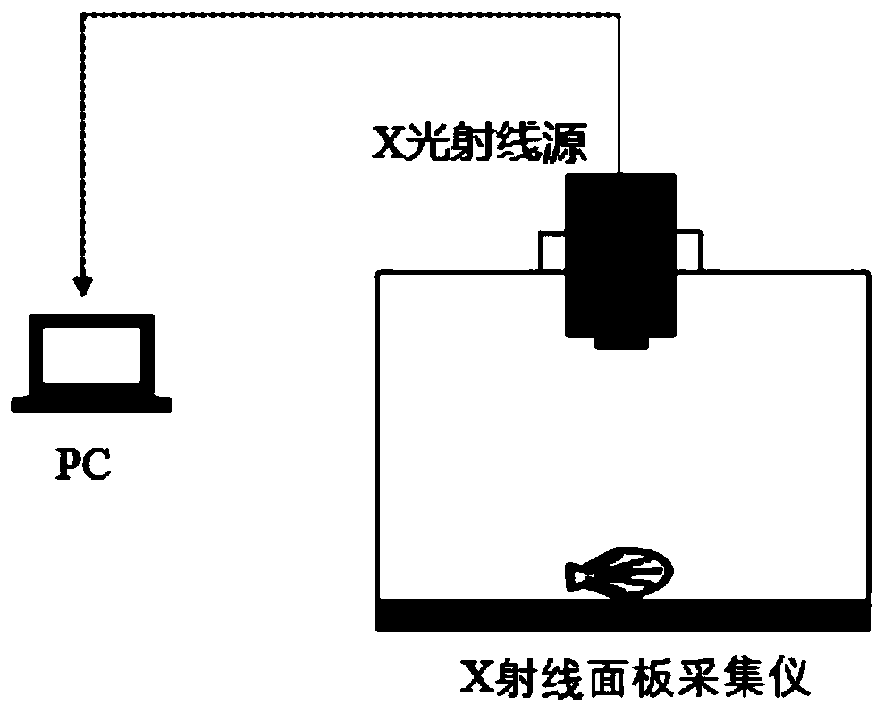
[0027] (2) Install the X-ray image acquisition device (manufacturer: Shenzhen Tianhe Times Electronic Equipment Co., Ltd. AT2310), specifically the X-ray source, X-ray panel acquisition instrument (including X-ray control box) and the X-ray acquisition device installed The PC side of the system software is connected together, such as figure 1 shown;
[0028] (3) Align the emission source port of the X-ray source with the "+" position (center position) of the X-ray panel collector, and the distance between the X-ray emission source and the panel of the collector is 1.2m-1.5m;
[0029] ...
Embodiment 2
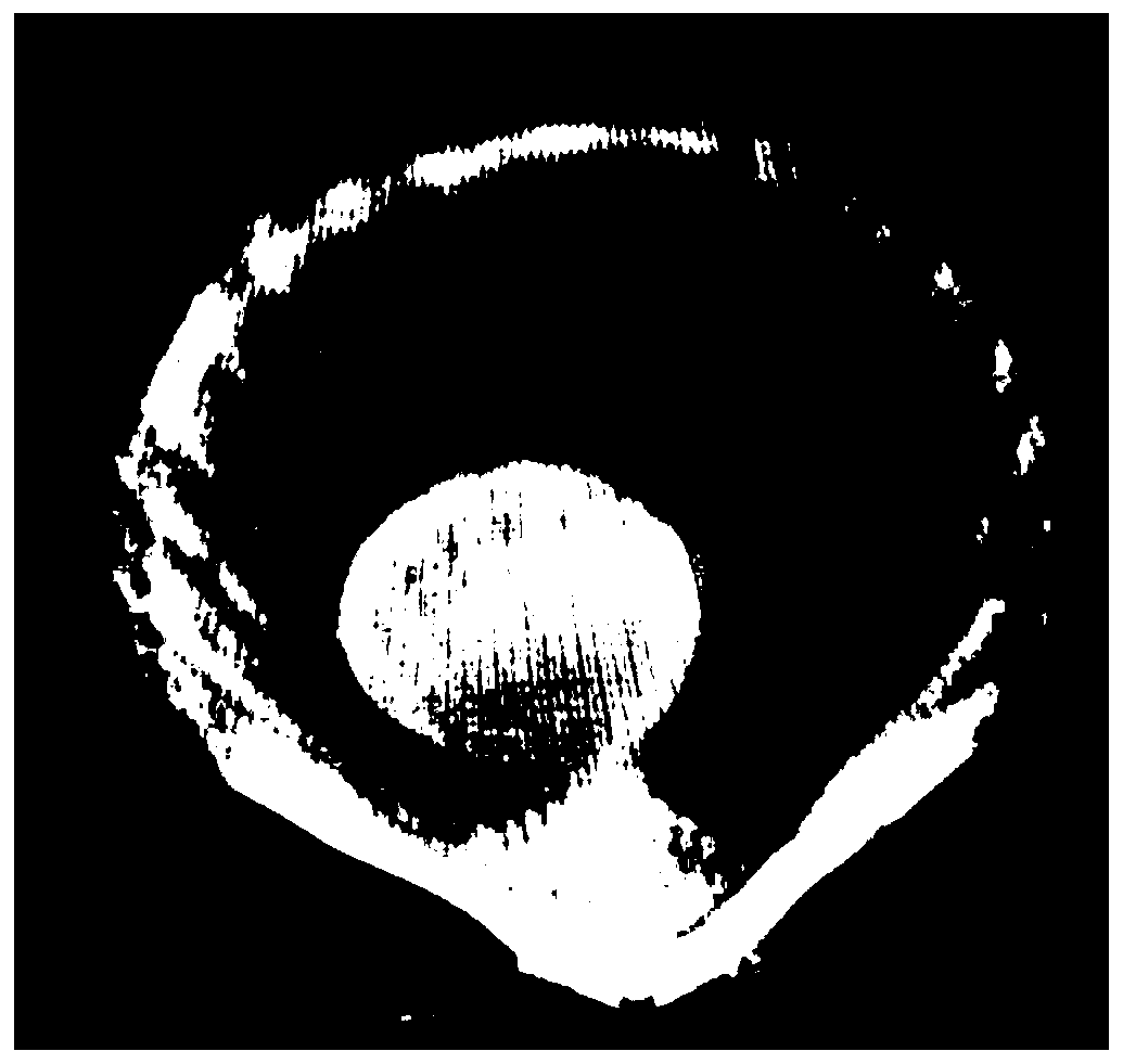
[0037] According to the following two settings, the X-ray image acquisition device is used to collect the X-ray image of the same scallop. The results are as follows: Figure 5 As shown (the first setting is on the left, the second setting is on the right). It can be seen from the figure that when the shell imaging effect is similar, the image of the adductor muscle in the image obtained by the first setting is clear, which can detect the size information of the adductor muscle more accurately.
[0038] First setting:
[0039] Radiation source parameter setting: high voltage value 120 (KV), current 800uA;
[0040] X-ray panel acquisition instrument parameter setting: select the software starting mode, set according to the following parameters: ExposureTime 1500ms (irradiation time 1500s), Delay Time 1000ms (delay time 1000s), Integrate Time 100ms (integration time 100ms), double exposure (secondary exposure) ;Click Apply to confirm the implementation of this parameter.
[004...
Embodiment 3
[0045] A total of 232 Ezo scallops of different ages and different sizes (the height of the first-year-old shell about 3cm, and the height of the third-year-old shell about 9cm) were selected, and a total of 232 pieces were used. The cross-sectional area of the adductor muscle was measured and counted by the method described in Example 1. The weight of the adductor muscle was measured in anatomically related scallops, and the correlation between the cross-sectional area of the scallops and the weight of the real meat column was calculated (Table 1). Significant correlation indicates that the area data of the scallop adductor muscle obtained under the non-destructive condition by using the present invention can be used to reflect the real weight, thereby indicating that the cross-sectional area can be used as a standard for shellfish species selection.
[0046] Table 1 Correlation of adductor muscle weight in scallops of different ages
[0047] One-ling clam S...
PUM
 Abstract
Description
Claims
Application Information
 - R&D
- Intellectual Property
- Life Sciences
- Materials
- Tech Scout
- Unparalleled Data Quality
- Higher Quality Content
- 60% Fewer Hallucinations
Browse by: Latest US Patents, China's latest patents, Technical Efficacy Thesaurus, Application Domain, Technology Topic, Popular Technical Reports.
© 2025 PatSnap. All rights reserved.Legal|Privacy policy|Modern Slavery Act Transparency Statement|Sitemap|About US| Contact US: help@patsnap.com