In-vitro method for evaluating repair function by reconstructing normal human body three-dimensional skin model

A skin model, human body technology, applied in the direction of material inspection products, measuring devices, instruments, etc., can solve problems such as evaluation methods or standards that have not proposed functions or efficacy, and achieve the effect of increasing credibility and high correlation

Inactive Publication Date: 2020-03-31
GUANGZHOU HUADAI BIOLOGICAL TECH CO LTD
View PDF1 Cites 4 Cited by
  • Summary
  • Abstract
  • Description
  • Claims
  • Application Information

AI Technical Summary

Problems solved by technology

However, the current international standards only focus on the safety evaluation of

Method used


Image

  • In-vitro method for evaluating repair function by reconstructing normal human body three-dimensional skin model
  • In-vitro method for evaluating repair function by reconstructing normal human body three-dimensional skin model
  • In-vitro method for evaluating repair function by reconstructing normal human body three-dimensional skin model

Examples


Embodiment 1

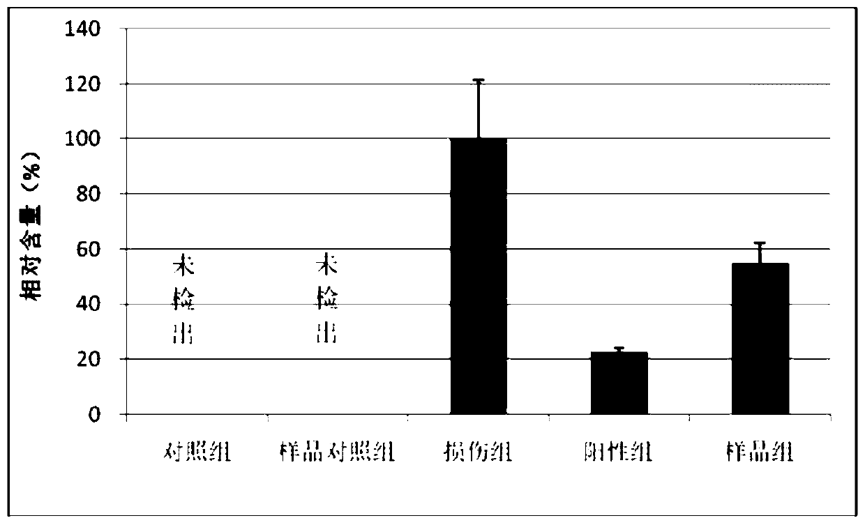
[0044] Example 1 Chitosan repair efficacy evaluation based on three-dimensional skin tissue

[0045] 1. Preparation for the experiment

[0046] 1.1 Recombinant human epidermis model kit: purchased commercially (purchased from Shanghai Sianpinuo Biotechnology Co., Ltd., Episkin TM detection kit), equipped with a dedicated maintenance medium.

[0047] 1.2 Sample preparation: Chitosan, as it is.

[0048] 1.3 Other reagents: SDS, PBS, MTT, EGF, isopropanol, ELISA kit.

[0049] 1.4 Incubation conditions: temperature 37±1°C, 5% CO 2 concentration, saturated relative humidity.

[0050] 1.5 Exposure conditions: room temperature, temperature 20-25°C, relative humidity 40-70%.

[0051] 2 Test process:

[0052] 2.1 Preparation of recombinant human skin model.

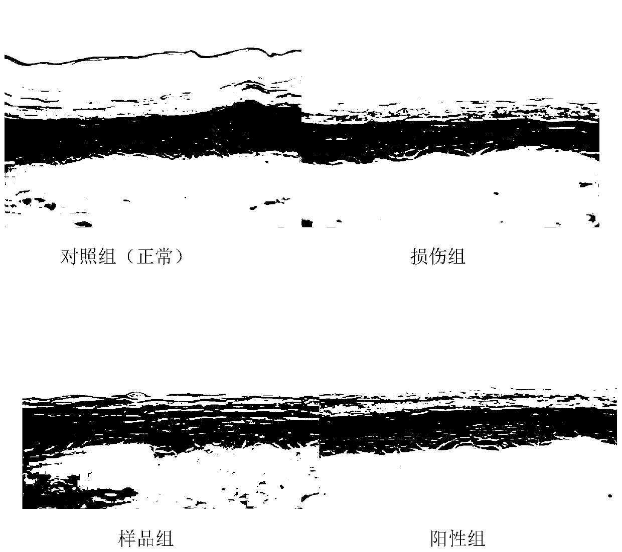
[0053] 2.1.1 Check the model and discard any visible damage or abnormality.

[0054] 2.1.2 Transfer the model to a culture plate containing 2mL of maintenance medium and incubate overnight.

[0055] 2.2 Test grouping and ...

Embodiment 2

[0085] Example 2 In vitro evaluation of the skin repair function of a basement membrane extract

[0086] 1. Preparation for the experiment

[0087] 1.1 Recombinant human epidermis model kit: purchased commercially (purchased from Shanghai Sianpinuo Biotechnology Co., Ltd., Episkin TM detection kit), equipped with a dedicated maintenance medium.

[0088] 1.2 Sample preparation: basement membrane extract

[0089] 1.3 Other reagents: PBS, MTT, EGF, isopropanol, tissue fixative, hematoxylin-eosin staining solution.

[0090] 1.4 Incubation conditions: temperature 37±1°C, 5% CO 2 concentration, saturated relative humidity.

[0091] 1.5 Exposure conditions: room temperature, temperature 20-25°C, relative humidity 40-70%.

[0092] 2 test process

[0093] 2.1 Preparation of recombinant human skin model

[0094] 2.1.1 Check the model, which meets the quality requirements of the model, and the skin resistance is 60-80Ω, indicating that the skin is not damaged and the barrier funct...

Embodiment 3

[0119] Example 3 Research on skin repair efficacy of trehalose and vitamin E based on three-dimensional skin tissue

[0120] 1. Preparation for the experiment

[0121] 1.1 Recombinant human epidermis model kit: purchased commercially (purchased from Shanghai Sianpinuo Biotechnology Co., Ltd., Episkin TM detection kit), equipped with a special culture medium.

[0122] 1.2 Sample preparation: trehalose and vitamin E

[0123] 1.3 Other reagents: PBS, MTT, fetal bovine serum, isopropanol, IL-1α and TNFα ELISA kits.

[0124] 1.4 Incubation conditions: temperature 37±1°C, 5% CO 2 concentration, saturated relative humidity.

[0125] 1.5 Exposure conditions: room temperature, temperature 20-25°C, relative humidity 40-70%.

[0126] 2 test process

[0127] 2.1 Preparation of recombinant human skin model

[0128] 2.1.1 Check the model and discard any visible damage or abnormality.

[0129] 2.1.2 Transfer the model to a 6-well culture plate containing 1 mL of maintenance medium an...


PUM


Abstract

The invention discloses an in-vitro method for evaluating a repair function by reconstructing a normal human body three-dimensional skin model. According to the method, an in-vitro recombined three-dimensional normal human skin model is utilized, SDS or a mechanical method is adopted to cause damage to the skin, the repairing effect of a tested substance on the damaged skin is evaluated by analyzing the recovery situation after damage or the intensity of inflammatory reaction, and the method specifically comprises the following steps: (1) preparation of a damage model, (2) restoration, (3) measurement of evaluation indexes, and (4) result analysis. The method has the advantages of high flux, repeatability, high correlation with people, multi-index evaluation and the like, and can be used for quickly evaluating various tested objects.

Description

technical field [0001] The invention belongs to the technical field of repair function evaluation, and in particular relates to an in vitro method for evaluating repair function by reconstructing a three-dimensional skin model of a normal human body. Background technique [0002] Skin covers the surface of the human body and is the outermost barrier between the human body and the perception and contact with the external environment. After the skin is damaged, an inflammatory reaction occurs early, manifested as redness, swelling, exudate, and formation of scabs. Subsequent proliferation of keratinocytes and fibroblasts, myofibroblast differentiation and wound contraction is followed by cellular and extracellular matrix structural remodeling and possible formation of granulation tissue or scarring. [0003] After the skin is damaged, physical, chemical and biological factors in the external environment directly contact human tissues, which can easily cause infection, chronic...

Claims


Application Information

Patent Timeline
IPC IPC(8): G01N33/68
CPCG01N33/6869G01N33/6863
Inventor 程树军黄健聪冯鉴鸿
Owner GUANGZHOU HUADAI BIOLOGICAL TECH CO LTD
Why Eureka
Try Eureka