Plasma molecular marker combination for predicting radiation damage

A technology for radiation damage and plasma, which is applied in the field of plasma molecular marker combinations for predicting radiation damage, and can solve problems such as large fluctuations and difficult radiation doses

Pending Publication Date: 2020-05-19
ACADEMY OF MILITARY MEDICAL SCI
View PDF0 Cites 3 Cited by
  • Summary
  • Abstract
  • Description
  • Claims
  • Application Information

AI Technical Summary

Problems solved by technology

However, if these metabolites do not have a dose-effect relationship with the radiation dose, do not have a time-effect relationship with the radiation time, or the expression levels of these metabolites fluctuate g...

Method used


Image

  • Plasma molecular marker combination for predicting radiation damage
  • Plasma molecular marker combination for predicting radiation damage
  • Plasma molecular marker combination for predicting radiation damage

Examples


Embodiment 1

[0075] Example 1. With the increase of irradiation dose, the degree of injury of mice gradually increased

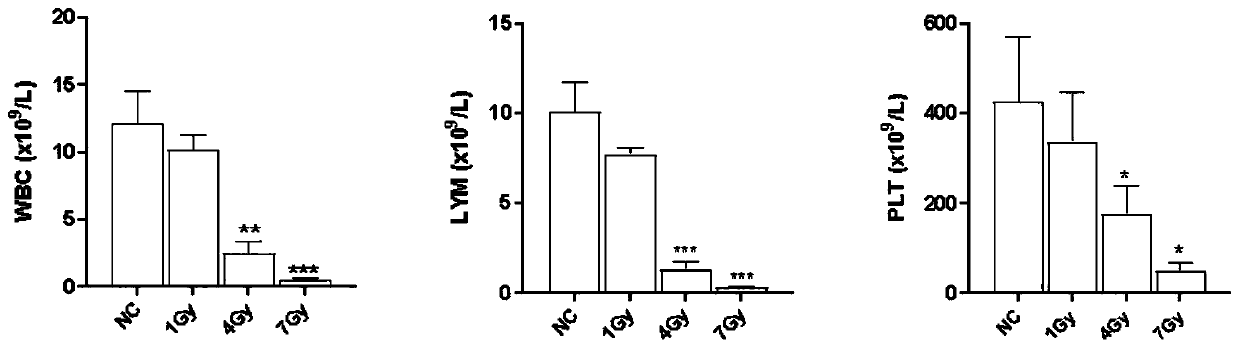
[0076] 6-8 weeks C57BL / 6 mice, a total of 40, were randomly divided into 4 groups, 10 mice in each group, and were subjected to 0, 1, 4, 7Gy 60 After Coγ-ray irradiation, blood was collected through the tail vein on the third day, and the number of white blood cells (WBC), lymphocytes (LYM) and platelets (PLT) in peripheral blood was analyzed with an automatic blood cell analyzer.

[0077] The result is as figure 1 shown. The results showed that after different doses of γ-ray irradiation, the number of WBC, LYM and PLT in the peripheral blood images gradually decreased with the increase of the irradiation dose, indicating that with the increase of the irradiation dose, the degree of injury of the mice was gradually aggravated.

Embodiment 2

[0078] Example 2. Preparation of samples for detection of plasma markers after irradiation in mice

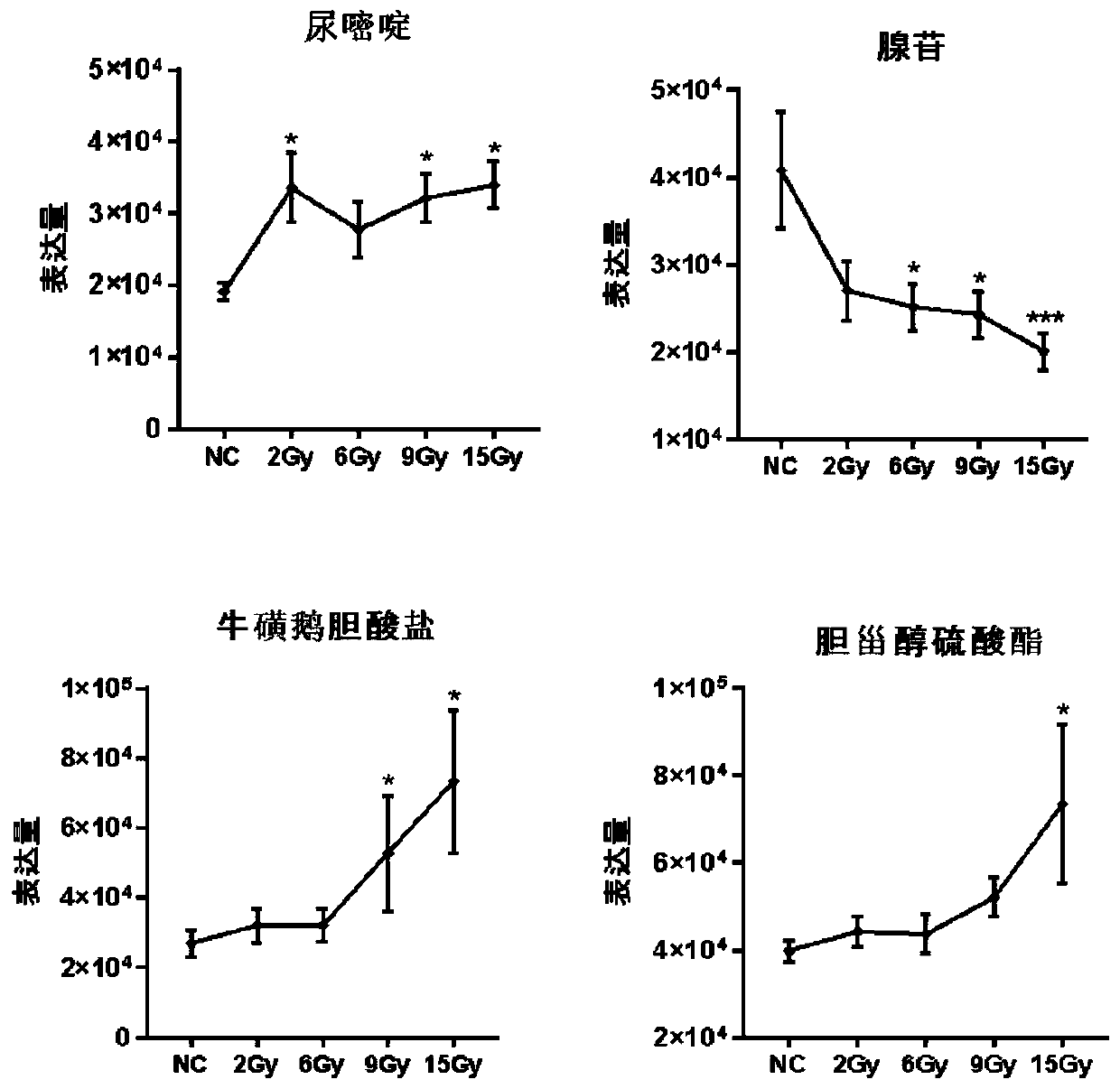
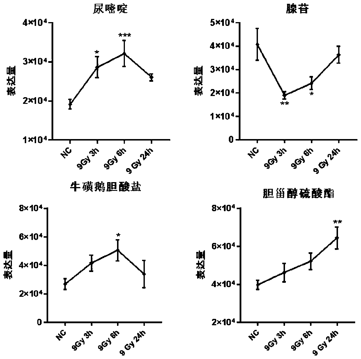
[0079] 6-8 weeks C57BL / 6 mice, a total of 80, were randomly divided into 8 groups, 10 mice in each group, and 5 groups were treated with 0, 2, 6, 9, 15Gy 60 Coγ-rays were irradiated, and blood was collected 6 hours after irradiation; the other three groups received 9Gy 60 Coγ-rays were irradiated, and blood was collected 3, 6, and 24 hours after irradiation.

[0080] After the mice were anesthetized, blood was collected from the heart, anticoagulated with heparin, centrifuged at 3000rpm, plasma was extracted, stored and transported at -80°C.

Embodiment 3

[0081] Embodiment 3, mass spectrometry sample processing and analysis

[0082] Take out the samples stored in Example 2 at -80°C, slowly dissolve at 4°C, take 100 μl of samples from each group, add 400 μl of pre-cooled methanol-acetonitrile solution (1:1, v / v), vortex for 60 seconds, and place at -20°C Precipitate protein for 1h, centrifuge at 14000rcf, 4°C for 20min, and take the supernatant to freeze-dry. The samples were separated by Agilent 1290 Infinity LC ultra-high performance liquid chromatography system (UHPLC) HILIC column, and detected by electrospray ionization (ESI) positive ion and negative ion modes respectively. The samples were separated by UHPLC and analyzed by mass spectrometry with Triple TOF 5600 mass spectrometer (AB SCIEX).

[0083] The specific experimental conditions are:

[0084] Chromatographic conditions: the samples were separated by Agilent 1290 Infinity LC ultra-high performance liquid chromatography system (UHPLC) HILIC column. The column t...


PUM


Abstract

The invention discloses a plasma molecular marker combination for predicting radiation damage. The invention provides application of eight target objects (uracil, adenosine, taurochenocholate, cholesteryl sulfate, linoleic acid, mono-2-ethylhexyl phthalate, thymine and 2-deoxyuracil) as biomarkers in the following steps, preparing a product for evaluating a radiation exposure dose, or evaluating the radiation exposure dose; and preparing a product for evaluating the radiation damage condition. The marker combination is advantaged in that mass spectrometry is used for detecting the level of small molecules in specific plasma, and that the eight target objects can serve as plasma biomolecular markers for predicting the radiation dose and the radiation damage degree is found. The method is ofgreat significance for quickly evaluating the irradiated dose and the radiation damage degree of large-scale irradiated crowds in the irradiated early stage after nuclear explosion or nuclear accidents so as to perform classified rescue and treatment and determine a treatment scheme.

Description

technical field [0001] The invention relates to the field of biotechnology, in particular to a combination of plasma molecular markers for predicting radiation damage. Background technique [0002] Mild to moderate ionizing radiation can cause serious damage to the human body, and severe ionizing radiation can cause death of humans or animals. In order to carry out targeted treatment quickly and accurately, it is necessary to identify those who are suspected of being exposed to radiation as early as possible. Assessment of exposure dose or degree of injury for classified rescue and treatment. At present, physical or biological methods are mostly used to evaluate the physical dose of radiation. Biological methods such as micronucleus detection methods are often time-consuming and require professionals to perform detection operations. In addition, because the environment in which the human body is exposed to radiation is different, the timing of exposure and the position of e...

Claims


Application Information

Patent Timeline
IPC IPC(8): G01N33/53G01N33/96G01N30/88
CPCG01N30/88G01N33/5308G01N33/96G01N2030/8822G01N2800/40
Inventor 葛常辉郑晓飞付汉江朱捷苏菲于海月
Owner ACADEMY OF MILITARY MEDICAL SCI
Why Eureka
Try Eureka