Construction method and application of organoid virus infection model
A construction method and virus infection technology, applied in the fields of biomedicine and cell engineering, can solve the problems of long cycle, unfavorable management, pathogenic microorganism contamination, etc.
- Summary
- Abstract
- Description
- Claims
- Application Information
AI Technical Summary
Problems solved by technology
Method used
Image
Examples
Embodiment 1
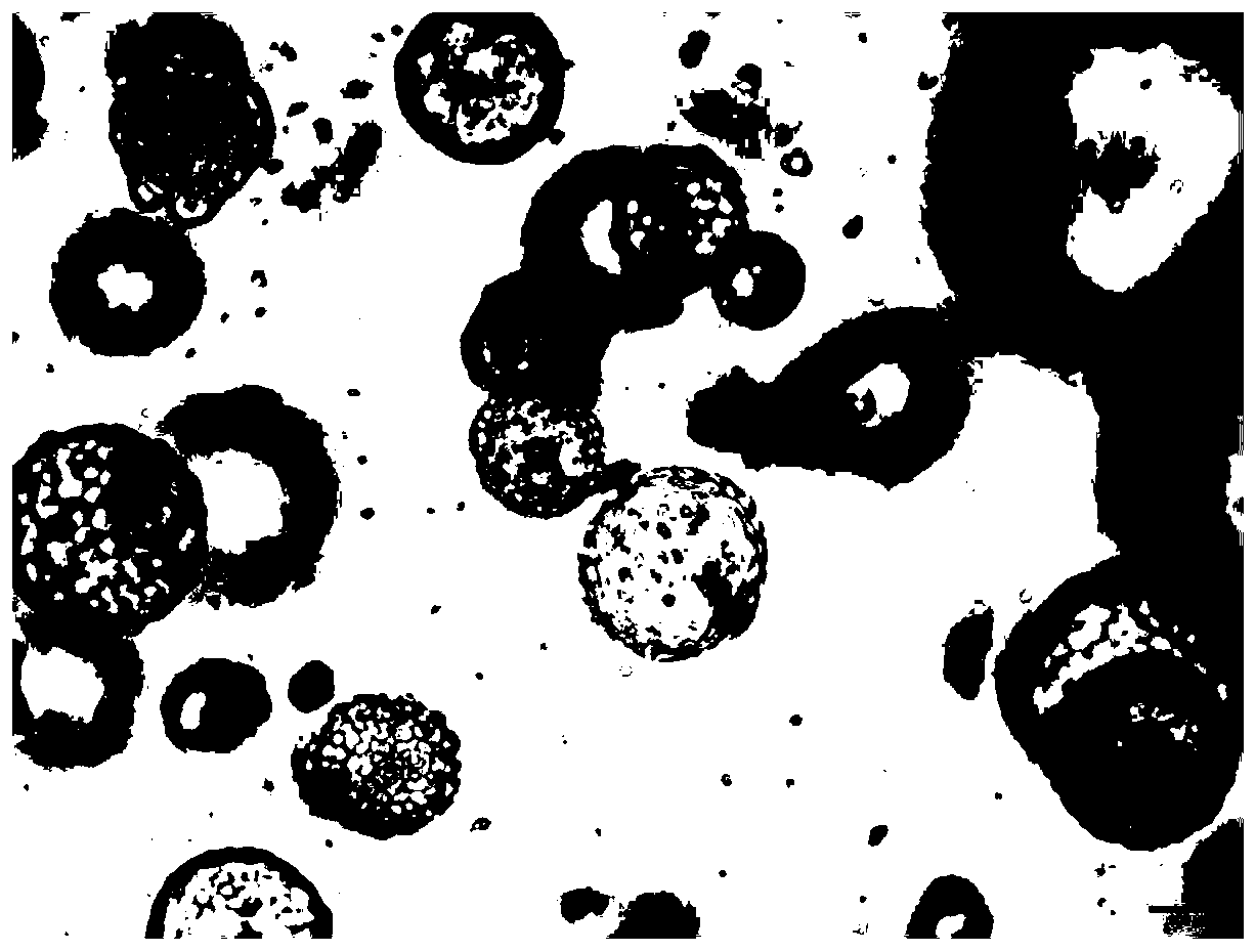
[0109] Example 1: Isolation and culture of human lung organoids
[0110] 1. Take the lung tissue resected by lung surgery and place it in the pre-cooled basal medium at 4°C. The basal medium components are: Advanced DMEM / F12 medium, penicillin, GlutaMax and HEPES to maintain the tissue cells. active.
[0111] 2. Cut the lung tissue obtained in step 1 into 0.5mm with surgical scissors 3 and transferred to a 15mL centrifuge tube, adding 10mL of washing medium (composition: DMEM medium, 1% fetal bovine serum, 1% penicillin) and repeatedly pipetting to obtain small pieces of lung tissue.
[0112] 3. Let the small pieces of lung tissue obtained in step 2 settle down, and remove the supernatant. Then add 10 mL of washing medium, mix by pipetting, and obtain small pieces of lung tissue.
[0113] 4. Remove the excess washing medium as much as possible from the small pieces of lung tissue obtained in step 3, and add 37°C preheated digestion medium, the composition of which is isolat...
Embodiment 2
[0132] Example 2: Isolation and culture of human liver organoids
[0133] 1. Take the liver tissue resected by liver surgery and place it in the pre-cooled basal medium at 4°C. The basal medium components are: Advanced DMEM / F12 medium, penicillin, GlutaMax and HEPES to maintain the tissue cells. active. , to obtain small pieces of liver tissue
[0134] 2. Cut the liver tissue obtained in step 1 into 0.5mm with surgical scissors 3 and transferred to a 15mL centrifuge tube, adding 10mL washing medium (composition: DMEM medium, 1% fetal bovine serum, 1% penicillin) and blowing repeatedly.
[0135] 3. Let the small pieces of lung tissue obtained in step 2 settle down, and remove the supernatant. Then add 10 mL of washing medium, mix by pipetting, and obtain small pieces of liver tissue.
[0136] 4. Remove as much excess washing medium as possible from the small pieces of lung tissue obtained in step 3, and add 37°C preheated digestion medium, the composition of which is collag...
Embodiment 3
[0152] Example 3: Isolation and culture of human intestinal organoids
[0153] 1. Take the intestinal tissue resected by intestinal surgery and place it in the pre-cooled basal medium at 4°C. The basal medium components are: Advanced DMEM / F12 medium, penicillin, GlutaMax and HEPES to maintain the tissue cell activity.
[0154] 2. Use a scalpel to gently scrape off the fluff on the inner wall of the intestinal tissue obtained in step 1, and then use surgical scissors to cut it into 0.5cm 3 and transfer it to a 15mL centrifuge tube, add 10mL PBS, and pipette repeatedly to obtain small pieces of intestinal tissue.
[0155] 3. Set aside the small pieces of intestinal tissue obtained in step 2 to settle, and remove the supernatant. Add 10 mL of PBS solution containing 5 mM EDTA and let stand at 4° C. for 25 minutes for digestion.
[0156] 4. Transfer the intestinal tissue digested in step 3 to a new Petri dish containing 10 mL of PBS, repeatedly pipet the intestinal tissue about...
PUM
 Abstract
Description
Claims
Application Information
 - R&D
- Intellectual Property
- Life Sciences
- Materials
- Tech Scout
- Unparalleled Data Quality
- Higher Quality Content
- 60% Fewer Hallucinations
Browse by: Latest US Patents, China's latest patents, Technical Efficacy Thesaurus, Application Domain, Technology Topic, Popular Technical Reports.
© 2025 PatSnap. All rights reserved.Legal|Privacy policy|Modern Slavery Act Transparency Statement|Sitemap|About US| Contact US: help@patsnap.com


