Intravascular plug-in visual flexible optical fiber surgical tool

A surgical tool and plug-in technology, applied in the fields of endoscopy, laparoscopy, medical science, etc., can solve the problems of inapplicable vascular imaging, etc., and achieve the effect of low cost, guaranteed surgical quality, and high robustness

Pending Publication Date: 2020-09-01
GUILIN UNIV OF ELECTRONIC TECH
View PDF4 Cites 9 Cited by
  • Summary
  • Abstract
  • Description
  • Claims
  • Application Information

AI Technical Summary

Problems solved by technology

[0007] Although the above-mentioned imaging system can be used for imaging transmission of cells, it requires additional lighting sources and is not suitable for imaging of blood vessels and in vivo

Method used


Image

  • Intravascular plug-in visual flexible optical fiber surgical tool
  • Intravascular plug-in visual flexible optical fiber surgical tool
  • Intravascular plug-in visual flexible optical fiber surgical tool

Examples


Embodiment 1

[0036] Example 1: Endovascular thrombus ablation surgery.

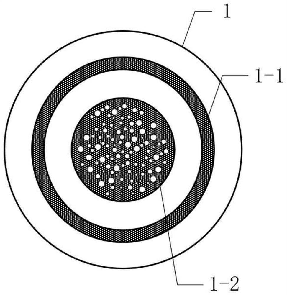
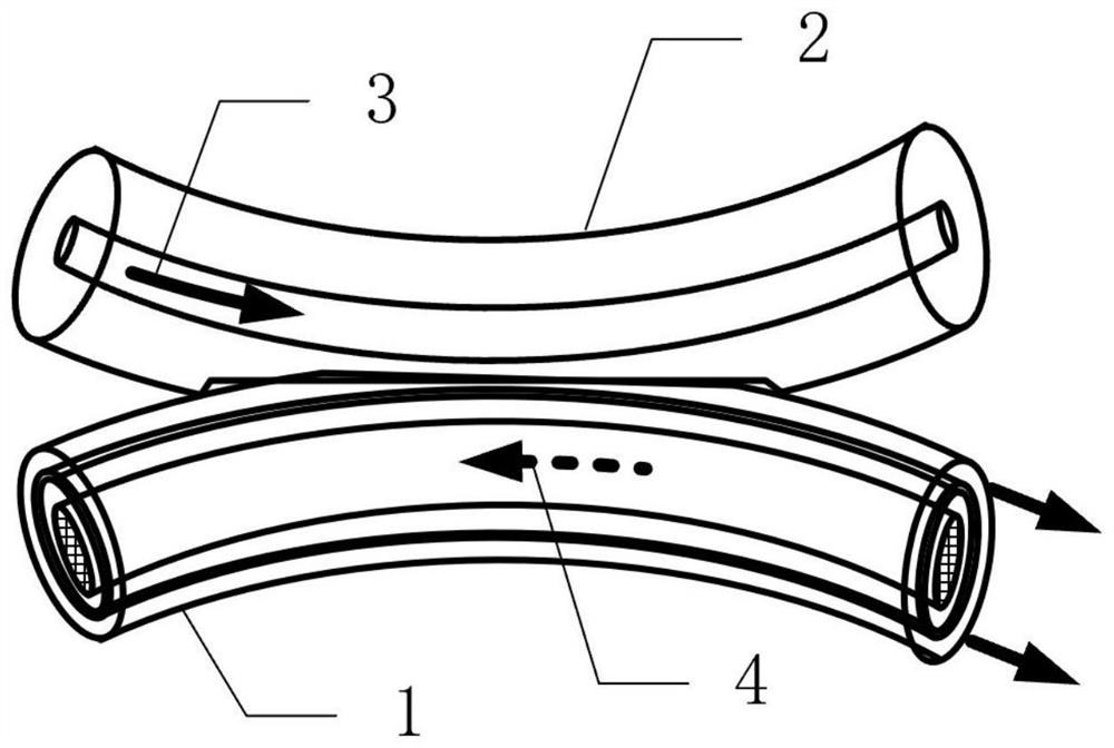
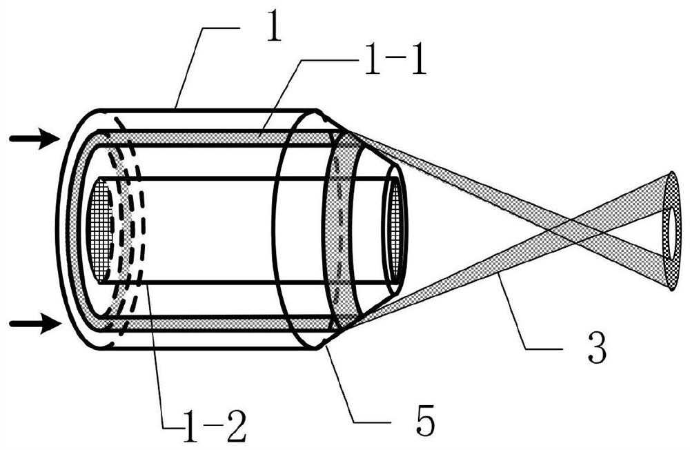
[0037] This embodiment adopts as figure 1 The Anderson Local Area Fiber 1 with ring waveguide is shown, the whole system is shown as Figure 4 shown. The system includes an incoherent broadband illumination source 7 , a power-adjustable surgical laser source 8 , a fiber wavelength division multiplexer 9 , a fiber side throw coupler 6 , an Anderson local fiber 1 with a ring waveguide, and a camera system 10 .

[0038] Among them, the illumination light source 7 adopts an LED light source with a center wavelength of 460nm; the surgical laser light source 8 selects an infrared semiconductor laser with a wavelength of 810nm, and the power is adjustable from 0 to 3W; the camera system uses a CCD detector, and there is an infrared cut-off filter in front of the detector to Prevent the surgical laser from reaching the detector and affect the imaging effect.

[0039] First, the fiber optic probe 1 proposed by the present i...

Embodiment 2

[0043] Example 2: Intravascular thrombus ablation surgery based on fluorescence imaging.

[0044] The difference between this embodiment and embodiment 1 is:

[0045] (1) In this embodiment, a certain amount of fluorescent marker needs to be injected into the blood vessel in advance, and the operation is performed after the thrombus region of the lesion specifically absorbs the fluorescent marker.

[0046] (2) The illumination light source used in this embodiment is a laser in the fluorescence excitation band of the fluorescent marker. After the cells in the diseased area absorb the fluorescently labeled drug, turn on the fluorescent excitation lighting source, and only the lesioned thrombus area in the blood vessel will emit fluorescence. Fluorescent images of the lesion area. Thrombectomy is then performed using a surgical light source.


PUM


Abstract

The invention provides an intravascular plug-in visual flexible optical fiber surgical tool. The intravascular plug-in visual flexible optical fiber surgical tool is characterized by consisting of anAnderson local optical fiber with an annular waveguide, a lighting source, a surgical light source, an optical fiber wavelength division multiplexer, an optical fiber lateral-fling coupler and a camera system. In the composition, (1) light waves of the lighting source and the surgical light source pass through the optical fiber wavelength division multiplexer and the optical fiber lateral-fling coupler and then are input into an annular core of the Anderson local optical fiber with an annular waveguide for transmission; (2) intravascular illumination is provided by an illumination beam transmitted in the annular waveguide, intravascular image information is collected by an Anderson local optical waveguide channel of the Anderson local optical fiber with the annular waveguide, the intravascular image information is transmitted to the camera system for detection, and a lesion area is searched; and (3) the lesion area in the blood vessel is removed by the operation light beam transmittedin the annular waveguide. The intravascular plug-in visual flexible optical fiber surgical tool can be used for imaging and operation of lesion areas such as tumors or thrombi in blood vessels, and can be widely applied to the field of cardiovascular disease treatment.

Description

[0001] (1) Technical field [0002] The invention relates to an intravascular insertion type visualized flexible optical fiber surgical tool, which belongs to the technical field of medical instruments. [0003] (2) Background technology [0004] At present, arterial cannulation technology has been very mature. Under the guidance of clinical imaging medicine, this technology inserts special catheters, guide wires and other tiny instruments into the lesion for diagnostic imaging and treatment through percutaneous puncture of blood vessels or the original channels of the human body. This technique is simple to operate, less invasive, and does not require suturing blood vessels. It completely replaces the previous method of surgical incision and exposure of blood vessels, and has become the basic operation technique of modern interventional therapy. Prevention, chemotherapy and other aspects have achieved good results. However, because the treatment is limited by the limitations...

Claims


Application Information

Patent Timeline
Patent Type & Authority Applications(China)
IPC IPC(8): A61B1/313A61B1/07A61B1/04
CPCA61B1/3137A61B1/07A61B1/0676A61B1/0684A61B1/042A61B1/043
Inventor 苑立波杨世泰
Owner GUILIN UNIV OF ELECTRONIC TECH
Why Eureka
Try Eureka