Biological tissue clearing reagent and biological tissue clearing method

A technology of biological tissue and reagents, applied in the preparation of test samples, material excitation analysis, fluorescence/phosphorescence, etc., can solve the problems of fluorescent protein fluorescence signal quenching, loss of biomolecules, low degree of transparency, etc., to improve transparency Speed ​​and effect, good imaging effect, effect of increasing permeability

Active Publication Date: 2021-12-14
UNIV OF SCI & TECH OF CHINA
View PDF0 Cites 0 Cited by
  • Summary
  • Abstract
  • Description
  • Claims
  • Application Information

AI Technical Summary

Problems solved by technology

[0004] Based on the above technologies, the SeeDB technology is generally low in transparency and requires multiple replacements of solutions with different gradients. Although the 3DISCO method has a high degree of transparency, the use of toxic organic solvents in the transparency requires careful operation and will cause Apparent quenching of the fluorescent signal of the fluorescent protein
CUBIC technology uses non-toxic reagents for tissue clearing to achieve a good clearing effect. However, this method uses a high concentration of detergent, which will cause a large number of biomolecules in the sample to be lost during the clearing process, resulting in fluorescence massive loss of signal
CLARITY uses toxic acrylamide for tissue cross-linking, which can preserve the biomolecules in the tissue well, but it needs to be handled carefully, and this method needs to completely remove the lipids in the tissue, which will lead to the loss of lipid signal markers, and The operation is complex and requires high experimental skills

Method used


Image

  • Biological tissue clearing reagent and biological tissue clearing method
  • Biological tissue clearing reagent and biological tissue clearing method
  • Biological tissue clearing reagent and biological tissue clearing method

Examples


Embodiment 1

[0044] A) fixation and separation of brain tissue, the embodiment all adopts mouse brain, and its fixation and separation method are as follows:

[0045] Prepare 1x PBS and pre-cooled 4% PFA at 4 °C. Mice were anesthetized with 10% chloral hydrate, and cardiac perfusion was performed after the mice were completely anesthetized. The perfusion process was as follows: first perfuse 20ml 1×PBS, then perfuse 20ml pre-cooled 4% PFA, and the perfusion speed was 10ml / min. Afterwards, the brain tissue was obtained by craniotomy. The sign of successful perfusion was that the brain was completely white without red blood vessels. The isolated brain was fixed in 4% paraformaldehyde at 4°C for 24h.

[0046] B) The brain tissue was cut into slices with a thickness of 300 μm using a vibrating microtome, and the slices were washed with 1×PBS for 1 hour. The tissue sections were then placed in PBS, 20% sorbitol (A), 20% dimethyl sulfoxide (B), 6% tromethamine (C), 40% 2,2′-thiodiethanol (D ...

Embodiment 2

[0049] A) Fixation and separation of brain tissue. The brain of a Thy1-YFP transgenic mouse was used in the embodiment, and the steps of fixing and separation of the brain were the same as those in Example 1.

[0050] B) The brain tissue was cut into 100 μm thick brain slices with a vibrating microtome, then placed in 1×PBS and washed for 10 minutes, sealed with PBS, and then imaged with an Olympus FV1200 laser confocal microscope. The imaging results are as follows figure 2 left picture.

[0051] C) Remove the imaged brain slice and put it into the clearing reagent, seal the slice with the clearing reagent after 5 minutes, and then use the Olympus FV1200 laser confocal microscope to image again with the same parameters and the same position. The imaging results are as follows figure 2 right picture. The fluorescence intensity was normalized with the data before clearing, and through statistical analysis, it was shown that the fluorescent signal of YFP was significantly enh...

Embodiment 3

[0053] A) Fixation and separation of brain tissue. The brain of a GAD-GFP transgenic mouse was used in the embodiment, and the steps of fixing and separation of the brain were the same as in Example 1.

[0054] B) The brain tissue was cut into 100 μm thick brain slices with a vibrating microtome, washed in 1×PBS for 10 min, mounted with PBS, and then imaged with an Olympus FV1200 laser confocal microscope.

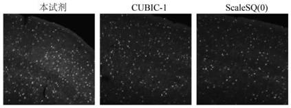
[0055] C) Remove the imaged brain slices and put them into common rapid tissue clearing reagents such as CUBIC-1, ScaleSQ(0) and the clearing reagent in Example 1 (containing 20% ​​sorbitol, 20% dimethyl sulfoxide , 40% 2,2′-thiodiethanol and 6% trometamol solution), after 5 minutes, use the corresponding clearing reagent to seal the slide, and use the Olympus FV1200 laser confocal microscope again with the same parameters and Imaging at the same position, the imaging results are as follows Figure 4 . Fluorescence intensity is carried out homogenization with the data be...


PUM

PropertyMeasurementUnit
thicknessaaaaaaaaaa

Abstract

The invention provides a biological tissue clearing reagent, comprising sorbitol, dimethyl sulfoxide, 2,2'-thiodiethanol and distilled water; the mass volume ratio of the sorbitol to distilled water is 15% to 25% ; The volume ratio of dimethyl sulfoxide to distilled water is 15%-25%; the volume ratio of 2,2'-thiodiethanol to distilled water is 40%-55%. Compared with the prior art, the present invention uses 2,2'-thiodiethanol as the main transparent component, adjusts the refractive index of the transparent reagent, and uses sorbitol to change the quaternary structure of the protein to produce a transparent effect. Dimethyl sulfoxide increases the permeability of tissues, improves the speed and effect of clearing, so that the obtained biological tissue clearing reagent can realize tissue clearing by simply processing biological samples while preserving and enhancing fluorescent labeling signals for better results. Imaging effect, wide applicability.

Description

technical field [0001] The invention belongs to the field of biological tissue sample processing, in particular to a biological tissue clearing reagent and a biological tissue clearing method. Background technique [0002] Tissue clearing technology is to use a variety of chemical reagents to process biological tissue, so that the refractive index of biological tissue is uniform, and the transparency is increased, thereby improving the imaging depth of the optical microscope, and then performing three-dimensional reconstruction of the shape of biological samples. [0003] According to the specific implementation plan of the tissue transparency technology, the currently commonly used tissue transparency technology is mainly divided into four categories. The first type is the simple immersion type, and the representative method is the SeeDB method invented by Takeshi Imai’s laboratory in 2013 (Ke, M.T., S.Fujimoto, and T.Imai, Nature Neuroscience, 2013.16(8): p.1154-U246. ), ...

Claims


Application Information

Patent Timeline
Patent Type & Authority Patents(China)
IPC IPC(8): G01N1/30G01N21/84G01N21/64
CPCG01N1/30G01N21/84G01N21/6458G01N21/6428
Inventor 周江宁单庆红秦新娅
Owner UNIV OF SCI & TECH OF CHINA
Why Eureka
Try Eureka