3D printing bone tissue engineering stent with slow release and osteogenesis promoting functions and preparation method and application of 3D printing bone tissue engineering stent

A tissue engineering scaffold and 3D printing technology, applied in the field of biomedical materials, can solve the problems of slow healing of bone tissue and poor osteogenic performance

Active Publication Date: 2020-10-27
SICHUAN UNIV
View PDF5 Cites 3 Cited by
  • Summary
  • Abstract
  • Description
  • Claims
  • Application Information

AI Technical Summary

Problems solved by technology

Bone tissue has a typical three-dimensional porous structure, which is especially suitable for 3D printing technology. 3D printed bone tissue engineering scaffolds have been used clinically, but there are still some shortcomings, such as poor bone formation performance, which leads to slow healing of bone tissue after implantation.

Method used


Image

  • 3D printing bone tissue engineering stent with slow release and osteogenesis promoting functions and preparation method and application of 3D printing bone tissue engineering stent
  • 3D printing bone tissue engineering stent with slow release and osteogenesis promoting functions and preparation method and application of 3D printing bone tissue engineering stent

Examples


Embodiment 1

[0047] (1) Preparation of 3D printing ink

[0048] In a water bath at 30°C, add sodium alginate powder material to deionized water to prepare a 6% sodium alginate solution, add 1ml of icariin-dimethyl sulfoxide solution with a concentration of 0.1g / ml, and use a magnetic Mix it well with a mixer until smooth. After preheating at 50°C for 30-60 minutes, reduce the viscosity of the sodium alginate solution, then slowly add the solid calcium phosphate powder into the preheated solution, and mix the slurry evenly with a homogeneous mixer until no phosphoric acid is observed Calcium agglomerates. The mass ratio of calcium phosphate, sodium alginate, icariin and deionized water in the final slurry is 25:6:0.08:100. This mixed slurry is used as 3D printing ink.

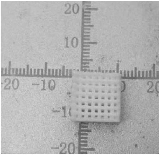
[0049] (2) printing support

[0050] The three-dimensional model of the scaffold was designed by 3D modeling software, and the mixed ink was printed into a calcium phosphate scaffold with a first-level macroporous structur...

Embodiment 2

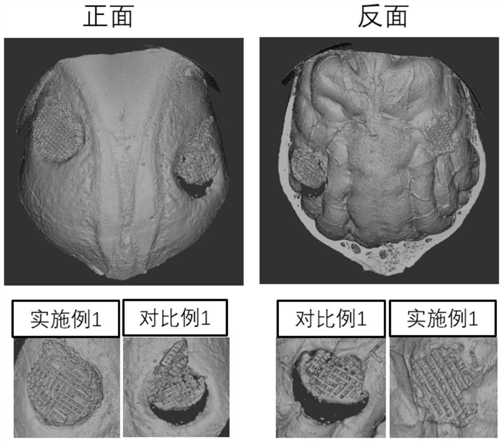
[0062] The 3D printed bone tissue engineering scaffold material was prepared with reference to the method described in Example 1, which differs from Example 1 in that the mass ratio of calcium phosphate, sodium alginate, icariin and deionized water was adjusted to 25:6 : 0.04: 100, all the other processing methods are the same as those described in Example 1. The experimental results show that the obtained scaffold has a slow-release function and an osteogenesis-promoting effect, but compared with Example 1, due to the lower solid content of icariin in the printing paste, bone tissue with slow-release and osteogenesis-promoting functions The total drug loading of the engineering scaffold is less, and the osteogenesis-promoting function of the scaffold obtained in this embodiment on bone defects is better than that of Comparative Example 1, but slightly worse than that of Example 1.

Embodiment 3

[0064] The 3D printed bone tissue engineering scaffold material was prepared with reference to the method described in Example 1, which differs from Example 1 in that the mass ratio of calcium phosphate, sodium alginate, icariin and deionized water was adjusted to 25:6 : 0.16: 100, all the other processing methods are the same as those described in Example 1. The experimental results show that the obtained scaffold has slow-release function and osteogenesis-promoting effect. Since the solid content of icariin in the printing slurry is more than that of Examples 1 and 2, the bone tissue engineering scaffold with slow-release and osteogenesis-promoting function The total amount of drug loaded is more, and the bone defect-promoting function of the scaffold obtained in this embodiment is better than that of Examples 1 and 2.


PUM

PropertyMeasurementUnit
concentrationaaaaaaaaaa

Abstract

The invention discloses a 3D printing bone tissue engineering stent with slow release and osteogenesis promoting functions and a preparation method of the 3D printing bone tissue engineering stent, and belongs to the technical field of biomedical materials. The stent is formed by in-situ crosslinking of a porous calcium phosphate material and sodium alginate, and is loaded with a drug, icariin. The bone tissue engineering stent prepared by the invention has good biocompatibility and bioactivity, and meanwhile, the osteogenic property of the stent is further enhanced by adding the drug icariininto printing ink; finally, a pore structure of the stent is regulated and controlled through 3D printing and a post-treatment mode, a crosslinking degree of the stent is regulated and controlled by changing concentration and crosslinking time of the calcium chloride crosslinking agent, and then an in-vivo degradation rate of the material is regulated and controlled. The drug sustained-release effect of the stent is achieved by changing the drug concentration to regulate and control the drug loading capacity of the stent, and the stent material can be applied to artificial bone and engineeringreconstruction and repair of bone tissue and has a wide application prospect clinically.

Description

technical field [0001] The invention belongs to the technical field of biomedical materials, and in particular relates to a 3D printed bone tissue engineering scaffold loaded with icariin for slow release to promote osteoinductive reconstruction and a preparation method thereof. [0002] technical background [0003] Bone tissue engineering scaffold is an important part of tissue engineering. It is to transplant the seed cells cultured in vitro onto the scaffold material, and then implant the material into the bone defect, supplemented by different measures to promote cell proliferation and differentiation, and achieve the purpose of repairing bone defect. . An ideal bone scaffold material should be able to simulate biological functions such as bone composition, structure, and mechanical properties, provide a temporary place for cell proliferation and differentiation, and guide cells and blood vessels to grow and multiply along the pores of the scaffold. Traditional bone sca...

Claims


Application Information

Patent Timeline
Patent Type & Authority Applications(China)
IPC IPC(8): A61L27/12A61L27/56A61L27/54A61L27/20A61L27/50A61L27/58B33Y10/00B33Y70/10B33Y80/00
CPCA61L27/12A61L27/20A61L27/50A61L27/54A61L27/56A61L27/58A61L2300/112A61L2300/23A61L2300/412A61L2300/602A61L2430/02B33Y10/00B33Y80/00B33Y70/10C08L5/04
Inventor 周长春张宸熙孙桓樊渝江梁洁雷皓远吴丽娜孙建勋肖玉梅蒋青张兴栋
Owner SICHUAN UNIV
Why Eureka
Try Eureka