Ultrasonic image left ventricular myocardium segmentation method and system and application

An ultrasound image and myocardial technology, applied in the field of image processing, can solve the problems of long time, time-consuming labeling of data, imbalance of positive and negative sample ratio, etc., to achieve the effect of strengthening boundary information

Active Publication Date: 2020-11-03
XIDIAN UNIV
View PDF25 Cites 1 Cited by
  • Summary
  • Abstract
  • Description
  • Claims
  • Application Information

AI Technical Summary

Problems solved by technology

[0005] (1) Existing technology to judge whether a patient has myocardial hypertrophy mainly relies on the rich clinical experience of doctors to find the boundary between the endocardium and epicardium to measure the thickness and see if the measured value meets the diagnostic criteria for hypertrophic cardiomyopathy. However, the repeatability of the process is poor, and due to different clinical experience, the measured thickness is different
[0006] (2) The traditional machine learning method first needs to manually extract features, but due to the large differences in the anatomical structure of the left ventricle of different HCM patients; and the traditional segmentation method such a

Method used


Image

  • Ultrasonic image left ventricular myocardium segmentation method and system and application
  • Ultrasonic image left ventricular myocardium segmentation method and system and application
  • Ultrasonic image left ventricular myocardium segmentation method and system and application

Examples


Embodiment Construction

[0069] In order to make the object, technical solution and advantages of the present invention more clear, the present invention will be further described in detail below in conjunction with the examples. It should be understood that the specific embodiments described here are only used to explain the present invention, not to limit the present invention.

[0070] Aiming at the problems existing in the prior art, the present invention provides a method, system and application for segmenting left ventricular myocardium in ultrasonic images. The present invention will be described in detail below with reference to the accompanying drawings.

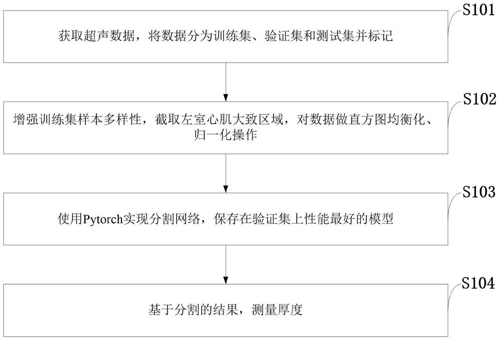
[0071] Such as figure 1 As shown, the segmentation method of the ultrasonic image left ventricular myocardium provided by the present invention comprises the following steps:

[0072] S101: Obtain ultrasound data, divide the data into training set, verification set and test set and mark them;

[0073] S102: Enhancing the diversity of samp...


PUM


Abstract

The invention belongs to the technical field of image processing, and discloses an ultrasonic image left ventricular myocardium segmentation method and system and an application. The method comprisesthe steps of obtaining ultrasonic data, dividing the data into a training set, a verification set and a test set, and performing marking; enhancing the diversity of training set samples, interceptingan approximate left ventricular myocardium region, and performing histogram equalization and normalization operation on the data; using Pytorch to realize network segmentation, and storing a model having the best performance on the verification set; and measuring the thickness based on a segmentation result. According to the ultrasonic image left ventricular myocardium segmentation method based ona convolutional neural network, the left ventricular myocardium at the end of diastole can be automatically segmented, shape information of the left ventricular myocardium is added into the network to assist network learning, proposed mixed loss functions are optimized from three angles, and boundary information is further enhanced during learning; and the thickness can be automatically measuredbased on the segmentation result, and no post-processing is needed in the whole process.

Description

technical field [0001] The invention belongs to the technical field of image processing, and in particular relates to a method, system and application for segmenting left ventricular myocardium in an ultrasonic image. Background technique [0002] At present, hypertrophic cardiomyopathy (HCM) is a common autosomal dominant cardiovascular disease, with an incidence of about 1:500-1:200 in the population and a mortality rate of about 1.4%-2.2%. , can lead to serious consequences such as chest tightness, chest pain, dyspnea, repeated syncope, atrial fibrillation, ventricular tachycardia, heart failure and even sudden death, and is the most common cause of sudden death in young people and athletes. At present, the main diagnostic criteria for HCM is left ventricular wall hypertrophy, which usually refers to interventricular septum or left ventricular wall thickness ≥ 15 mm measured by two-dimensional echocardiography, or thickness ≥ 13 mm in patients with a clear family history,...

Claims


Application Information

Patent Timeline
IPC IPC(8): A61B8/08G06N3/04G06T5/40G06T7/11
CPCA61B8/085A61B8/0858A61B8/5215G06T7/11G06T5/40G06T2207/30048G06N3/045
Inventor 任胜寒王永兵王倩胡芮赵恒刘丽文
Owner XIDIAN UNIV
Why Eureka
Try Eureka