Antibody, test strip and kit against bovine nodular skin disease virus
A technology for nodular and skin diseases, applied in the field of immunity, can solve the problems of expensive, complicated and time-consuming separation and culture identification methods
- Summary
- Abstract
- Description
- Claims
- Application Information
AI Technical Summary
Problems solved by technology
Method used
Image
Examples
Embodiment 1
[0034] Example 1: Prokaryotic expression and purification of P32 protein
[0035] In this example, the prokaryotic expression and purification of P32 protein are carried out, and the specific steps are as follows:
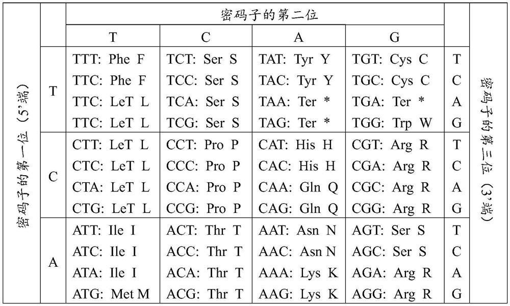
[0036] 1. Query the LSDV genome sequence from GeneBank, select the conserved gene LSDV-P32 through alignment analysis, and express the P32 protein. The amino acid sequence of P32 protein was analyzed by the online software TMHMM, and it was found that there was a transmembrane domain at the C-terminus. Because the transmembrane domain protein was insoluble, the C-terminal transmembrane domain was removed during expression and codon optimization was performed. The amino acid sequence of LSDV P32 protein is shown in Table 3.
[0037] Table 3: Amino acid sequence of P32 protein
[0038]
[0039] 2. Select the correctly sequenced pGEX-6P-2-N280 plasmid, transform it into BL21(DE3) competent cells, and cultivate overnight in a 37°C constant temperature incubator. ...
Embodiment 2
[0041] Example 2: Preparation of P32 protein monoclonal antibody
[0042] Eight 8-12-week-old female Balb / c mice were immunized with the P32 protein antigen purified in Example 1. After immunizing 3 times, the mice were collected orbital blood to obtain mouse serum, and ELISA was used to detect P32 in mouse serum. Protein antibody titer, if the titer is less than 10,000, boost immunization 1-2 times until the antibody titer is greater than 10,000.
[0043] The mouse splenocytes with antibody titer>10000 were fused with myeloma SP2 / 0 cells, and the fusion cells were screened by HAT selection medium, and the fusion cells were screened and subcloned by ELISA. Ascites, the antibody was purified by Protein A / G antibody purification column. The ELISA titer of the purified antibody was >1:128000, and the purity was >90%.
[0044] The cell line numbers corresponding to the two monoclonal antibodies are 701 and 901, respectively.
Embodiment 3
[0045] Example 3: Amplification and Sequence Determination of CDR Region Sequence of P32 Protein Monoclonal Antibody
[0046] The hybridoma cell lines 701 and 901 were recovered and cultured. When the cells grew to the logarithmic growth phase, the cell count was about 8×10. 7 cells / ml, cells were collected. Extract the total RNA of hybridoma cells according to the instructions of TaKaRa MiniBEST Universal RNA Extraction Kit, according to the reaction system shown in Table 4, incubate at 65 °C for 5 min, then quickly cool on ice, and then set the reaction system shown in Table 5 at 42 °C for 60 min; 70 ℃15min; 25℃1min for 1st-Strand cDNA synthesis reaction.
[0047] Table 4: 1st-Strand cDNA synthesis reaction mix
[0048] reagent Usage amount Oligo dT Primer (50μM) 1μL dNTP Mixture (10mM each) 1μL template RNA <5μg
RNase Free dH 2 O
Up to 10μL
[0049] Table 5: Reverse Transcription Reaction Solution
[0050] reag...
PUM
| Property | Measurement | Unit |
|---|---|---|
| particle diameter | aaaaa | aaaaa |
Abstract
Description
Claims
Application Information
 - R&D
- Intellectual Property
- Life Sciences
- Materials
- Tech Scout
- Unparalleled Data Quality
- Higher Quality Content
- 60% Fewer Hallucinations
Browse by: Latest US Patents, China's latest patents, Technical Efficacy Thesaurus, Application Domain, Technology Topic, Popular Technical Reports.
© 2025 PatSnap. All rights reserved.Legal|Privacy policy|Modern Slavery Act Transparency Statement|Sitemap|About US| Contact US: help@patsnap.com


