Lymph node classification method, system and device based on multi-view semi-supervision

A classification method and technology of lymph nodes, applied in the field of medical image processing, can solve the problems of difficulty in obtaining, limiting effective image information extraction, different background information, etc., and achieve the effect of improving accuracy

Pending Publication Date: 2021-03-19
XI AN JIAOTONG UNIV
View PDF4 Cites 5 Cited by
  • Summary
  • Abstract
  • Description
  • Claims
  • Application Information

AI Technical Summary

Problems solved by technology

The features obtained by this type of method are mostly traditional gray-scale-based image features or medical features that require fine calibration. Usually, the descriptive information obtained by traditional methods is relatively simple, which is not enough to extract sufficient and effective features for gray-scale ultrasound images; and Fine medical feature calibration requires calibration by professional doctors, which is usually not easy to obtain; these limitations limit the extraction of effective information from images
The development of deep learning in the field of image analysis has made data-driven methods successful in the field of medical imaging. However, the current existing methods usuall

Method used


Image

  • Lymph node classification method, system and device based on multi-view semi-supervision
  • Lymph node classification method, system and device based on multi-view semi-supervision
  • Lymph node classification method, system and device based on multi-view semi-supervision

Examples


Embodiment Construction

[0037] The present invention is described in further detail below in conjunction with accompanying drawing:

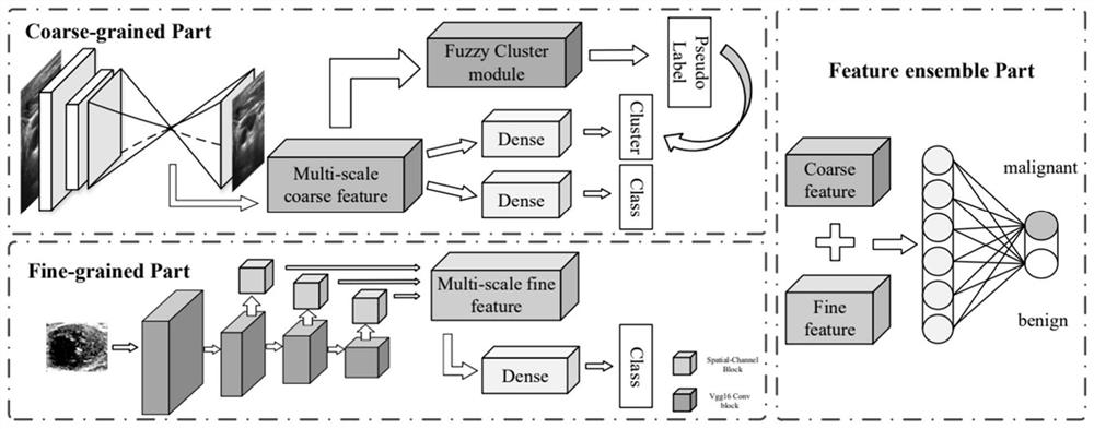
[0038] Such as figure 1 As shown, a multi-view semi-supervised lymph node classification method includes the following steps:

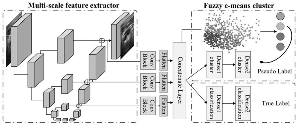
[0039] S1: Perform image preprocessing on the original gray-scale ultrasound image (coarse-grained image) of lymph nodes, and use image reconstruction neural network (Hourglass network) to perform image reconstruction on the preprocessed image;

[0040] The image reconstruction neural network includes an encoder and a decoder. The structure of the encoder and decoder includes a downsampling layer and an upsampling layer. When performing image reconstruction, downsampling is performed first to obtain the feature expression of the original image at different scales, and then upsampling is performed. Before each sampling, the residual module is used to operate, and in the process of upsampling, a cross-layer connection is added for point additio...


PUM


Abstract

The invention discloses a lymph node classification method, system and device based on multi-view semi-supervision. The method comprises the steps: carrying out the image preprocessing of an originalgray-scale ultrasonic image of a lymph node, carrying out the image reconstruction of the preprocessed image through a U-shaped neural network, and carrying out the weighted fusion to obtain a multi-scale fused coarse-grained image feature; performing coarse-grained feature representation learning in a semi-supervised manner; the method comprises the following steps of: cutting an ROI (Region Of Interest) region containing nodules of an original gray-scale ultrasonic image to obtain a fine-grained image, performing weighting processing on the fine-grained image through a vgg16 network in whicha space and channel attention mechanism is added at different levels, performing global average pooling, and splicing feature outputs at different levels to obtain fine-grained fusion features; the multi-view information is obtained by fusing the coarse-grained view features and the fine-grained attempt features, so that the fused features can have nodule environment information and detail information at the same time, richer and more accurate description is obtained, accurate classification can be carried out, and the classification accuracy is improved.

Description

technical field [0001] The invention belongs to the field of medical image processing, and in particular relates to a classification method, system and equipment for lymph nodes based on multi-view semi-supervision. Background technique [0002] Clinically, fine needle aspiration biopsy (FNA) is usually used as the gold standard for lymph node identification. Although it can provide accurate results, invasive examination and even surgery may lead to cervical lymph node lesions and affect the patient's physical condition to a certain extent . As a non-invasive method, ultrasound has become the most commonly used method for preoperative information collection of cervical lymph nodes due to its convenience and economy. The metastatic lymph nodes of thyroid cancer have certain ultrasound characteristics, which can be used to analyze the preoperative cervical lymph node metastasis of differentiated thyroid cancer patients and provide reference for lymph node dissection. Cervica...

Claims


Application Information

Patent Timeline
IPC IPC(8): G06T7/00G06K9/46G06K9/62G06N3/04G06N3/08
CPCG06T7/0012G06N3/08G06T2207/10132G06T2207/20081G06T2207/20084G06T2207/20104G06T2207/30004G06V10/40G06N3/045G06F18/23G06F18/253G06F18/24
Inventor 辛景民罗怡文刘思杰
Owner XI AN JIAOTONG UNIV
Why Eureka
Try Eureka