PDE6B nucleotide sequence and use thereof

A nucleotide sequence and sequence technology, applied in the PDE6B nucleotide sequence and its application field, can solve the problems of transducing retinal tissue cells, etc., and achieve the effects of preventing or treating retinitis pigmentosa, increasing thickness, and strong stimulation response

Active Publication Date: 2021-03-26
WUHAN NEUROPHTH BIOTECHNOLOGY LTD CO
View PDF1 Cites 0 Cited by
  • Summary
  • Abstract
  • Description
  • Claims
  • Application Information

AI Technical Summary

Problems solved by technology

[0003] Naturally occurring AAV serotypes are generally unable to transduce retinal tissue cells on the side of the vitreous cavity due to the presence of barriers such as the inner limiting membrane, glial cells, etc. that prevent the spread of AAV virions

Method used


Image

  • PDE6B nucleotide sequence and use thereof
  • PDE6B nucleotide sequence and use thereof
  • PDE6B nucleotide sequence and use thereof

Examples


Embodiment 1

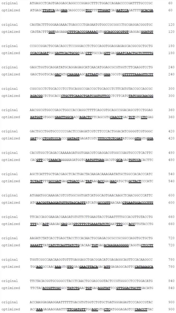
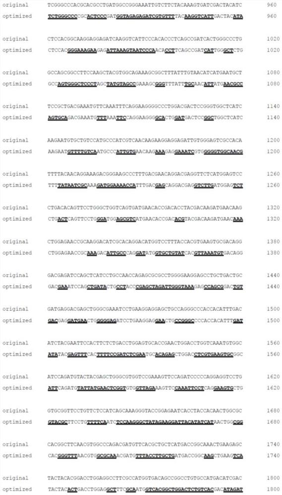
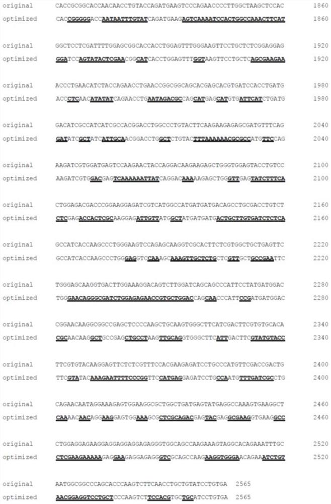
[0043] Example 1: Codon-optimized PDE6B vector construction and expression verification

[0044] (1) Plasmid vector construction

[0045] 1. Digest the backbone of the AAV2-CAG plasmid and the coPDE6B fragment or wtPDE6B fragment simultaneously with HindIII and XhoI respectively, and then ligate the digested fragments to the backbone respectively.

[0046] 2. The ligation product was transformed into E. coli, and a single colony was picked for enzyme digestion verification and sequencing verification.

[0047] (2) Cell transfection

[0048] 1.293 cells were plated in the cell culture well plate, and the cells were grown until the confluence reached 70-80%.

[0049] 2. Replace the medium with DMEM+1XGlutaMAX.

[0050] 3. Dilute the plasmid and PEI reagent with culture medium and mix well at a ratio of 1:2. After mixing, let stand at room temperature for 20 minutes, add the mixture to the cell culture medium, and shake gently.

[0051] 4. Place the cell culture plate in 37°C...

Embodiment 2

[0070] Example 2: CAG promoter has higher expression efficiency in rd10 mice

[0071] (1) Mice injected with virus drugs

[0072] 1. Prepare 5*10 12 vg / ml of AAV2 / 2.7m8-CAG-coPDE6B, AAV2 / 2.7m8-sCBA+AT2RIntron 1-coPDE6B and AAV2 / 2.7m8-sCBA-coPDE6B drugs.

[0073] 2. Inject 1 ul / eye of the above three viruses into the eyes of different age-appropriate rd10 mice respectively through the vitreous cavity or subretinal injection.

[0074] 3. At 3 weeks after the mice were injected, the mice were sacrificed, and the retinal tissues of the mice were separated.

[0075] (2) qPCR detection of PDE6B mRNA expression level

[0076] 1. Pre-cool the mortar with liquid nitrogen, add mouse eye tissue into the mortar and grind it into powder.

[0077] 2. Transfer the powder into an EP tube filled with Trizol lysate, shake vigorously and let stand at room temperature for 5 minutes, then centrifuge at 10,000 rpm and 4°C for 10 minutes.

[0078] 3. Transfer the supernatant to a new EP tube, a...

Embodiment 3

[0087] Example 3: AAV-coPDE6B gene therapy drug improves ocular function and repairs retinal structure in rd10 mice

[0088] (1) Mice injected with virus drugs

[0089] 1. Prepare 5*10 12 vg / ml of AAV2 / 2.7m8-CAG-coPDE6B drug and AAV2 / 2.7m8-CAG-GFP.

[0090] 2. Inject 1 ul / eye of AAV2 / 2.7m8-CAG-coPDE6B drug and AAV2 / 2.7m8-CAG-GFP virus into the eyes of age-appropriate mice through the vitreous cavity or subretinal injection.

[0091] 3. At 3 weeks after the mice were injected, the mice were sacrificed, and the retinal tissues of the mice were separated and made into slices for later use.

[0092] (2) Electroretinogram analysis

[0093] 1. The mice were anesthetized and the pupils were dilated, and at the same time, 2.5% hypromellose liquid containing electrodes was dripped into the eyes, and the corneal potential response was recorded.

[0094] 2. Under the condition of dark adaptation, let the mice adapt to the dark overnight, give short-term flash stimulation with differe...


PUM


Abstract

The invention relates to the technical field of biomedical gene therapy and discloses a PDE6B nucleotide sequence and use thereof. The protein expression efficiency of the codon-optimized PDE6B codingsequence is higher than that of a wild type sequence. AAV2/2.7 m8-coPDE6B drug treatment can obviously relieve retinal pathological symptoms of mice with retinal pigment degeneration caused by a PDE6B mutation and achieve a recovery of retinal functions. After the AAV2/2.7m8-coPDE6B drug is injected, the PDE6B can be efficiently expressed on a retinal outer nuclear layer and increase thickness ofthe retinal outer nuclear layer. A retina potential diagram shows that mice of an AAV2/2.7m8-coPDE6B drug treatment group has a strong stimulation response. Therefore, the AAV2/2.7m8-coPDE6B drug hasan effect of preventing or treating retinal pigment degeneration.

Description

technical field [0001] The present invention relates to the technical field of biomedical gene therapy, and more specifically relates to a PDE6B nucleotide sequence and its application. Background technique [0002] PDE6 protein consists of two catalytic subunits, PDE6A and PDE6B, and two inhibitory subunits. The protein can regulate the level of cGMP in the cytoplasm of the rod cells when they are stimulated by light, so that the rod cell membranes are hyperpolarized, and finally the receptor potential is generated. PDE6B mutation will lead to the inactivation of PDE6 protein, the destruction of light transduction pathway, the sharp increase of cGMP and calcium ion levels in the cytoplasm, causing apoptosis and degeneration of visual cells. Because PDE6B is indispensable in the light transduction pathway of rod cells, PDE6B gene mutations can cause severe retinitis pigmentosa, and there is currently no effective cure. [0003] Naturally occurring AAV serotypes are general...

Claims


Application Information

Patent Timeline
Patent Type & Authority Applications(China)
IPC IPC(8): C12N15/55C12N15/864A61K48/00A61K38/46A61P27/02
CPCC12N9/16C12N15/86A61K48/005A61K48/0008A61P27/02C12Y301/04035C12N2750/14143C12N2800/107C12N2800/22A61K38/00
Inventor 李斌任盛
Owner WUHAN NEUROPHTH BIOTECHNOLOGY LTD CO
Why Eureka
Try Eureka