Monomolecular protein detection chip and method based on ultramicro electrode array electrochemiluminescence

A protein detection and single-molecule technology, applied in single-molecule protein detection, integrated biomimetic microfluidic chip, and ultra-microelectrode array electrochemiluminescence, can solve the problems of chip failure, poor consistency and repeatability, and complicated device preparation, etc. question

Active Publication Date: 2021-04-16
SHENZHEN INST OF ADVANCED TECH
View PDF9 Cites 1 Cited by
  • Summary
  • Abstract
  • Description
  • Claims
  • Application Information

AI Technical Summary

Problems solved by technology

[0008] Among the single-molecule protein detection technologies that have been disclosed so far, there is no ultra-microelectrode array electrochemiluminescence-related technology. The main problems are: complex device preparation, unstable reaction process, and inconvenient effective control, which makes its detection consistency and poor repeatability
[0009] Among the currently published electrochemiluminescence immunoassay techniques, they mainly focus on the preparation and surface modification of three-electrode systems or bipolar electrodes, and the chip cannot detect trace protei...

Method used


Image

  • Monomolecular protein detection chip and method based on ultramicro electrode array electrochemiluminescence
  • Monomolecular protein detection chip and method based on ultramicro electrode array electrochemiluminescence
  • Monomolecular protein detection chip and method based on ultramicro electrode array electrochemiluminescence

Examples


Embodiment 1

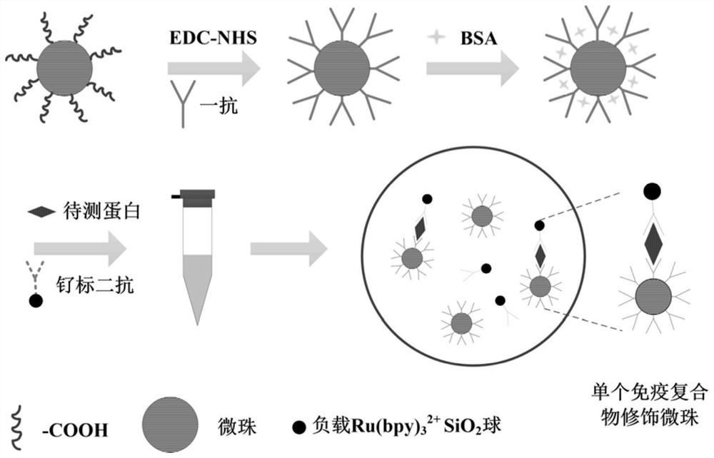
[0085] Example 1 Preparation of immune complex-modified microbeads and ruthenium-labeled functionalization

[0086] In order to ensure that each microbead binds at most one single-molecule protein and increase the intensity of electrochemiluminescence, an immune complex modified microbead and ruthenium-labeled functionalization method were designed. It mainly includes carboxylated microbeads activated by carbodiimide-N-hydroxysuccinimide (EDC-NHS) coupled with primary antibody, bovine serum albumin (BSA) blocks the immune active site, captures the target protein in the sample and Combined with ruthenium-labeled secondary antibody to form immune complex modified microbeads:

[0087] Specifically:

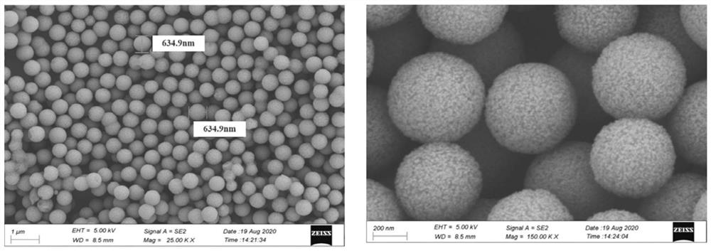
[0088] 1) 2.8 μm magnetic microbeads modified with carboxyl groups (10mL solution contains 6×10 8 ~7×10 8 microbeads) as the carrier to carry out sufficient carboxylation on the outside of the chip;

[0089] Prepare N-hydroxysuccinimide (NHS) with phosphate buffer (0.01M, pH 7.4)...

Embodiment 2

[0093] Example 2 Design and preparation of a single-molecule protein detection chip

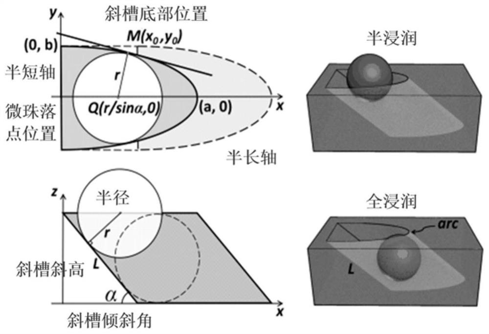
[0094] Design the specific structure of the single-molecule protein detection chip (see image 3 ): The basic microwell structural unit used to accommodate 2.8 μm microbeads is a semi-elliptical inclined groove microwell. axis, the inclination angle of the chute, etc., to construct the standard elliptic equation; the semi-major axis of the semi-ellipse is a, the semi-minor axis is b, and the inclination angle of the chute is α. According to the calculation, its size requirements need to meet the standard Ellipse equation:

[0095]

[0096] x and y are the coordinates of any point on the semi-ellipse, and the length of the semi-major axis of the semi-ellipse is a, then we can know a according to the formula 2 =4R 2 / sin 2 α, the length of the semi-minor axis is b, then it can be known that b is calculated according to the formula 2 =4R 2 The tilt angle of the / 3 tilted microwell is α,...

Embodiment 3

[0099] Embodiment 3 Research on the Specific Dimensions of the Microwell Array Unit

[0100] Firstly, it is determined that the SU-8 inclined microwell array prepared on the ITO electrode is 4000 rows × 20 columns, and the potential difference at both ends of a single microwell depends on the ratio of the length of the microwell to the distance between the two copper wires. When the potential difference is large enough, an oxidation or reduction reaction occurs on the electrode surfaces at both ends of the microwell, causing electrochemiluminescence. The highest potential is obtained when the drop point of the microbead is close to the semi-short axis. Therefore, the specific dimensions of the designed microwell array unit are 10 μm in the semi-major axis and 1.6 μm in the semi-minor axis. When an external voltage is applied across the ITO electrodes and a solution exists in the microwell array, a potential gradient is generated due to resistance. Because the resistance of t...


PUM

PropertyMeasurementUnit
Radiusaaaaaaaaaa
Particle sizeaaaaaaaaaa

Abstract

The invention relates to a monomolecular protein detection chip and a monomolecular protein detection method based on ultramicro electrode array electrochemiluminescence. The monomolecular protein detection chip is provided with a substrate of an ITO (indium tin oxide) electrode and an inclined micro trap array with a three-dimensional concave part on the ITO electrode, the two ends of the inclined micro trap array in the transverse direction are respectively provided with wires connected with the positive electrode and the negative electrode of a power supply. The single-molecule protein detection chip is simple to prepare, an electrochemical luminescence detection system further amplifies a signal, and high-sensitivity single-molecule detection of trace protein can be realized.

Description

technical field [0001] The present invention mainly relates to the fields of electroanalytical chemistry and microfluidics, and proposes a method for arraying microbeads on a microchip and an electrochemiluminescent method for matching ultramicroelectrode arrays, and develops a set of integrated bionic microfluidics accordingly. chip for single-molecule protein detection. The microfluidic chip obtains a fast and sensitive response signal by designing an ultra-microelectrode array, and secondly uses electrochemiluminescence to improve detection performance. It has the advantages of high detection sensitivity, short detection time, and low cost, and is especially suitable for single-molecule protein detection. potential economic value. Background technique [0002] With the continuous improvement of protein detection sensitivity requirements in the modern medical field, the development of advanced protein detection technology is the only way to improve the diagnostic ability ...

Claims


Application Information

Patent Timeline
IPC IPC(8): G01N27/416G01N33/68G01N33/552G01N33/543G01N33/532
Inventor 吴天准惠允
Owner SHENZHEN INST OF ADVANCED TECH
Why Eureka
Try Eureka