
Cell capture method based on micro-tube fluidic chip

A chip and cell technology, applied in the field of medical testing, can solve problems such as complicated operation, cumbersome steps, and expensive reagents

Active Publication Date: 2021-04-27
SHANGHAI JIAO TONG UNIV
View PDF12 Cites 0 Cited by
  • Summary
  • Abstract
  • Description
  • Claims
  • Application Information

AI Technical Summary

Problems solved by technology

[0006] In view of the defects of the existing separation technology that the steps are cumbersome, the reagents are expensive, and rare cells are easy to lose, a cell capture method based on a microtube fluidic chip is proposed. Utilize the effect of nucleated cells in the blood flow to the wall and the specific adhesion characteristics of cell antigens and antibodies to separate nucleated cells in whole blood, without the need to split red blood cells in advance or remove mature red blood cells by centrifugation, which solves the complicated operation of traditional separation methods. Time-consuming and other problems, with simple, convenient, fast and accurate and reliable characteristics

Method used


Image

  • Cell capture method based on micro-tube fluidic chip
  • Cell capture method based on micro-tube fluidic chip
  • Cell capture method based on micro-tube fluidic chip

Examples


Embodiment 1

[0035] This embodiment relates to the preparation operation of the micropipe fluidic chip, including:

[0036] 1) The polydimethylsiloxane (PDMS) matrix is ​​mixed evenly with the curing agent.

[0037] 2) Take the silicon plate, place the copper wire with a diameter of 100 μm, and keep the copper wire and the silicon plate at 0.34mm to tighten and fix the copper wire.

[0038] 3) Pouring PDMS to completely wrap the copper wire, and place it in a 60°C oven for two hours to cure.

[0039] 4) Take out the cured PDMS, and pull out the copper wire to obtain a PDMS microchannel fluidic chip with an inner diameter of 100 μm.

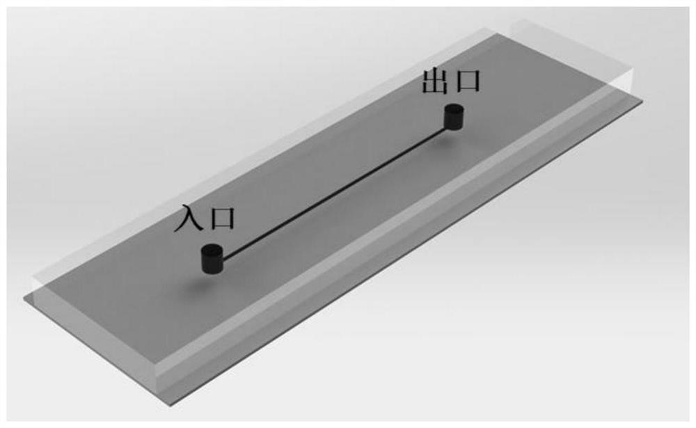
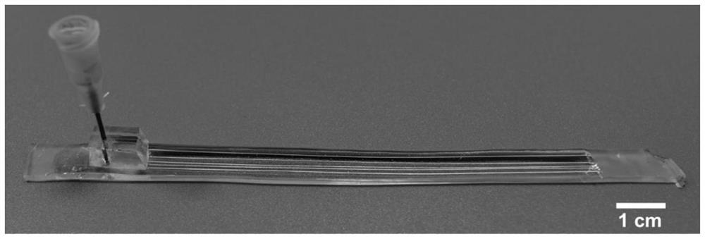
[0040] Such as figure 1 , figure 2 As shown, this embodiment relates to the microchannel fluidic chip prepared by the above method, that is, a PDMS plate including single or multiple circular channels.

[0041] This embodiment relates to the internal antibody modification and confirmation operations based on the above-mentioned microtubules, including:

...

Embodiment 2

[0048] This embodiment relates to the fabrication of a microtube chip for nucleated red blood cells, and the specific steps are:

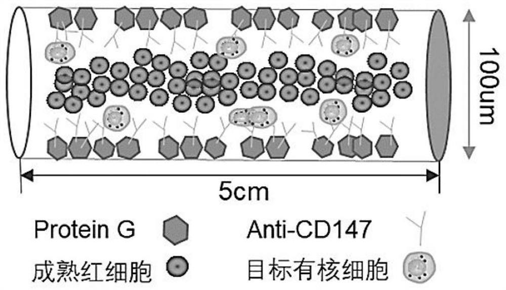
[0049] 1) Take the microtube chip prepared in Example 1, inject the IgG-conjugated CD147 antibody stock solution to fill the microtube, incubate overnight in a wet box at 4°C, and then wash the microtube with PBS to remove the unfirmly bound antibody.

[0050]2) In order to avoid non-specific adsorption of non-target cells, inject 2% fetal bovine serum albumin (BSA) to fill the microtubes, incubate in a humid box at room temperature for half an hour, and then rinse the microtubes with PBS; after the surface treatment of the microtubes, put them into 4°C refrigerator for standby to capture nucleated red blood cells.

[0051] This embodiment involves the sample processing and cell capture operation of the above-mentioned microtube chip, and the specific steps are:

[0052] 1) Pass 20 microliters of umbilical cord blood into the antibody-modified mic...

Embodiment 3

[0056] This embodiment relates to a method for in situ identification of fetal nucleated erythrocytes from umbilical cord blood in microtubes, and the specific steps are:

[0057] 1) Incubate with 0.05% glutaraldehyde to fix the cells, and wash the inner wall of the microtube with PBS after standing at room temperature for 10 minutes.

[0058] 2) Incubate with 0.01% Triton to perforate the cell membrane, and wash the inner wall of the microtube with PBS after standing at room temperature for 6 minutes.

[0059] 3) In order to avoid non-specific staining by the antibody, incubate with 2% BSA, let stand at room temperature for 6 minutes, and then wash the inner wall of the microtube with PBS.

[0060] 4) Incubate the HBF (hemoglobin F) + CD71 (PE) fluorescent antibody mixture, stain at 4°C overnight and wash the inner wall of the microtube with PBS to remove excess antibody in the tube.


PUM


Abstract

A cell capture method based on a micro-tube fluidic chip comprises the following steps: modifying G protein on the inner wall of a micron-diameter high-molecular polymer micro-tube through a chemical modification method, and then grafting an antibody with IgG, so that the tube wall adsorbs the antibody; enabling the sample to flow into a micro-tube fluidic chip, and identifying the captured target nucleated cells by utilizing an immunofluorescence technology. In the invention, nucleated cells in the whole blood are separated by utilizing the wall-approaching edge set effect of the nucleated cells in the blood flow and the specific adhesion characteristic of cell antigens and antibodies, and mature red blood cells do not need to be split in advance or centrifugally removed; therefore, the problems of complex operation, time consumption and the like of a traditional separation method are solved, and the method has the characteristics of simplicity, convenience, rapidness, accuracy and reliability.

Description

technical field [0001] The invention relates to a technique in the field of medical detection, in particular to a method for capturing cells from whole blood based on a microtube fluidic chip. Background technique [0002] Rare cells refer to cells with extremely low content in the human body, including fetal nucleated red blood cells in the peripheral blood of pregnant women, and circulating tumor cells in the peripheral blood of cancer patients. One hundred thousandth of a mature red blood cell. Rare cells have very low content in peripheral blood and are not easy to obtain, but they have important clinical significance. Traditional rare nucleated cell enrichment techniques are mainly divided into affinity enrichment and physical enrichment methods. [0003] Among them, the physical enrichment mainly depends on the special size, density and deformability of the target cells for separation. The more common ones are density gradient centrifugation (DGC), filtration based o...

Claims


Application Information

Patent Timeline
IPC IPC(8): G01N33/569G01N33/58G01N33/53
CPCG01N33/56966G01N33/582G01N33/5304G01N2469/10G01N2333/70503
Inventor 龚晓波刘洁张晟泓
Owner SHANGHAI JIAO TONG UNIV
Learn More