Injectable decellularized scaffold for cartilage repair as well as preparation method and application of injectable decellularized scaffold

A decellularized scaffold and cartilage repair technology, applied in the field of medical materials, can solve the problems of inability to simulate the multi-layer structure of cartilage tissue, poor fixation, slow release, etc.

Active Publication Date: 2021-05-18
GUANGDONG GENERAL HOSPITAL
View PDF5 Cites 6 Cited by
  • Summary
  • Abstract
  • Description
  • Claims
  • Application Information

AI Technical Summary

Problems solved by technology

However, KGN itself is not soluble in water, and the effect of direct injection is weak, so it is extremely important to find a suitable carrier to load KGN to achieve its slow release during the repair process
[0012] Cartilage tissue itself has a layered structure. In response to this characteristic, researchers have also developed a dual-phase scaffold material ...

Method used


Image

  • Injectable decellularized scaffold for cartilage repair as well as preparation method and application of injectable decellularized scaffold
  • Injectable decellularized scaffold for cartilage repair as well as preparation method and application of injectable decellularized scaffold

Examples


Embodiment 1

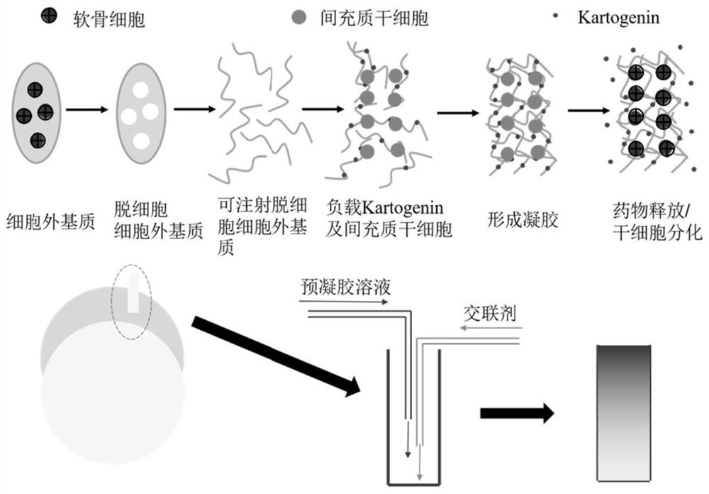
[0068] Step 1: Cartilage tissue was cut into 0.5mm thick slices, frozen at -80°C and ground into coarse powder. The powder was placed in a buffer solution of 1% emulsifier and stirred for 24 hours. Subsequently, the sample was placed in a mixed solution of DNase and RNase and stirred for 8 hours. Place it again in a buffer solution of 1% emulsifier and stir for 72 hours, and then soak in a large amount of deionized water overnight to remove residual chemical substances. Freeze at -80°C and continue to lyophilize, then grind into fine powder, and add to 15mg / mL pepsin / 0.01mol / L HCl solution. The mass ratio of pepsin to fine powder is 1:10. The resulting suspension was stirred at room temperature for 48 h for complete digestion. The resulting viscous solution was adjusted to neutrality using 0.1mol / L NaOH solution and 10×PBS in an ice-water bath to obtain injectable dECM;

[0069] Step 2: The synthesis of mesoporous silica was prepared by the improved Stober method adding po...

Embodiment 2

[0075] Step 1: Cartilage tissue was cut into 0.7mm thick slices, frozen at -80°C and ground into coarse powder. The powder was placed in a buffer solution of 1.5% emulsifier and stirred for 24 hours. Subsequently, the sample was placed in a mixed solution of DNase and RNase and stirred for 8 hours. Place it again in a buffer solution of 1% emulsifier and stir for 48 hours, and then soak in a large amount of deionized water overnight to remove residual chemical substances. Finally, the resulting dECM was frozen at -80°C and further lyophilized, then ground into a fine powder and added to a 15 mg / mL pepsin / 0.01 mol / L HCl solution. The mass ratio of pepsin to substrate is 1:2. The resulting suspension was stirred at room temperature for 48 h for complete digestion. The viscous solution was adjusted to neutrality using 0.1mol / L NaOH solution and 10×PBS in an ice-water bath to obtain injectable dECM;

[0076]Step 2: The synthesis of mesoporous silica was prepared by the improve...

Embodiment 3

[0082] Step 1: Cartilage tissue was cut into 0.7mm thick slices, frozen at -80°C and ground into coarse powder. The powder was placed in a buffer solution of 3% emulsifier and stirred for 24 hours. Subsequently, the sample was placed in a mixed solution of DNase and RNase and stirred for 12 hours. Place it again in a buffer solution of 3% emulsifier and stir for 48 hours, and then soak in a large amount of deionized water overnight to remove residual chemical substances. Finally, the resulting dECM was frozen at -80°C and further lyophilized, then ground into a fine powder and added to a 15 mg / mL pepsin / 0.01 mol / L HCl solution. The mass ratio of pepsin to substrate is 1:50. The resulting suspension was stirred at room temperature for 48 h for complete digestion. The viscous solution was adjusted to neutrality using 0.1mol / L NaOH solution and 10×PBS in an ice-water bath to obtain injectable dECM;

[0083] Step 2: The synthesis of mesoporous silica was prepared by the improv...


PUM

PropertyMeasurementUnit
Viscosityaaaaaaaaaa
Elastic modulusaaaaaaaaaa

Abstract

The invention discloses an injectable decellularized scaffold for cartilage repair as well as a preparation method and application of the injectable decellularized scaffold, and relates to the technical field of medical materials. According to the method, a double-injection mode is adopted, and the gelated gradient injectable decellularized scaffold is obtained by injecting a pre-gel solution and a viscosity modifier simultaneously. The decellularized scaffold can load MSCs to be implanted into a body, is hydrolyzed under a physiological pH value, and slowly releases a medicine at an injured part, so that the differentiation of the loaded MSCs to cartilage cells is promoted, and the injured cartilage tissue is repaired; Due to the gradient property, original biological characteristics of the cartilage tissue can be better simulated, and a better repairing effect is achieved. According to the decellularized scaffold and the technology, the raw materials are cheap and wide in source, after clinical popularization, the pain of a patient can be remarkably relieved, the operation frequency is reduced, the treatment effect is improved, and remarkable social and economic benefits are achieved.

Description

technical field [0001] The invention relates to the technical field of medical materials, in particular to an injectable decellularized scaffold for cartilage repair and its preparation method and application. Background technique [0002] The knee joint is one of the most structurally complex joints in the human body and plays a comprehensive and complex stabilizing role. In recent years, the national fitness movement has been gradually popularized and developed, coupled with my country's gradual aging society, the knee joint disease has shown a linear upward trend, especially cartilage damage has always been a common problem in clinical medicine. [0003] The clinical manifestations of cartilage damage are mainly joint pain, effusion and dysfunction. Cartilage damage is mainly common in repeated chronic injuries and degeneration. Once the best treatment time is delayed, it will easily lead to traumatic arthritis and further affect the patient's activity function. Knee ar...

Claims


Application Information

Patent Timeline
IPC IPC(8): A61L27/36A61L27/02A61L27/52A61L27/54A61L27/58
CPCA61L27/58A61L27/52A61L27/3687A61L27/3654A61L27/3633A61L27/54A61L27/025A61L2400/06A61L2430/06A61L2300/624A61L2300/21
Inventor 于啸天邓展涛郑秋坚马元琛
Owner GUANGDONG GENERAL HOSPITAL
Why Eureka
Try Eureka