Method and system for identifying epileptic focus of temporal lobe epilepsy caused by hippocampal sclerosis and/or predicting pathological typing of epilepsy
A technology for pathological typing and hippocampus, applied in character and pattern recognition, recognizing medical/anatomical patterns, image analysis, etc. question
- Summary
- Abstract
- Description
- Claims
- Application Information
AI Technical Summary
Problems solved by technology
Method used
Image
Examples
Embodiment 1
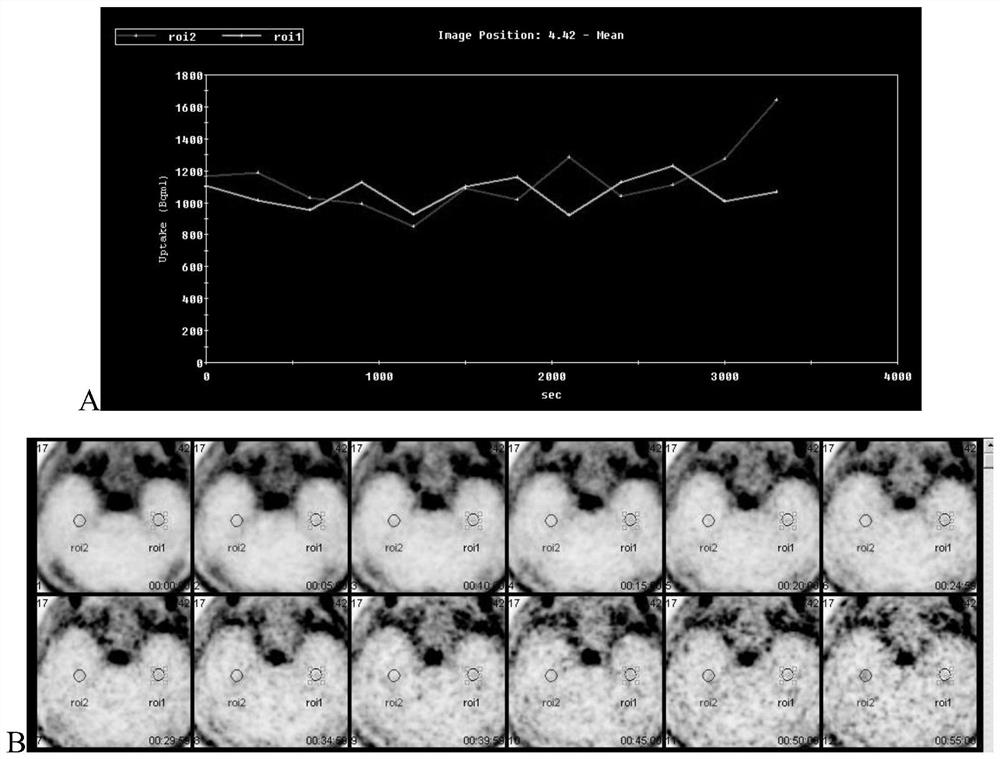
[0111] Example 1. Construction of time-radioactivity curve and metabolic characteristic fitting curve of positron radiopharmaceuticals
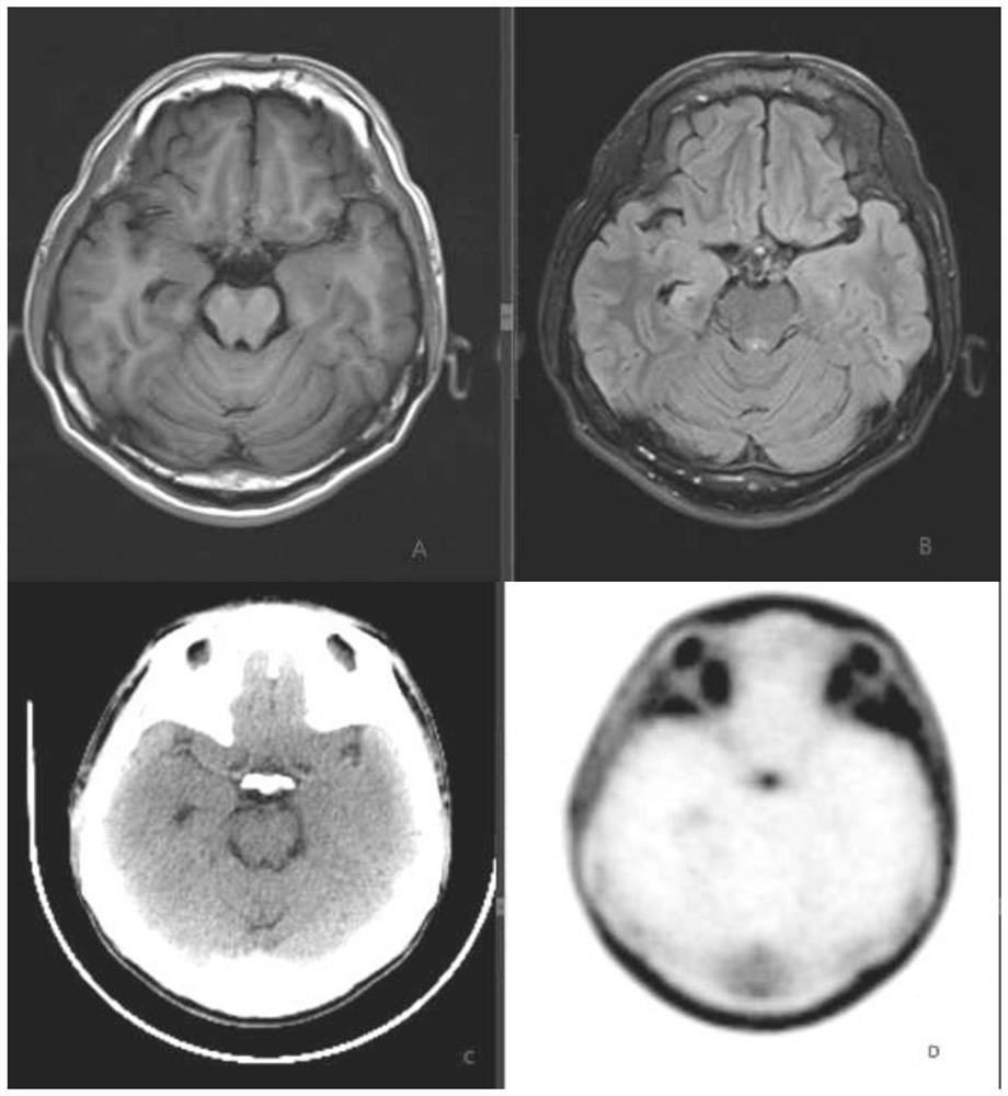
[0112] (1) Research objects: Patients with epilepsy caused by hippocampal sclerosis were selected for surgical treatment, and all patients underwent interictal treatment before surgery. 11 C-choline, 18 F-FDG, 11 C-FMZ PET (PET / CT or MR) brain imaging (three-day method).
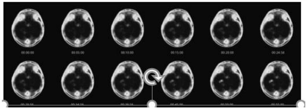
[0113] (2) Imaging conditions: 11 C-choline, 18 F-FDG, 11 C-FMZ uses 7.4MBq / kg, 5.55MBq / kg, and 5.55MBq / kg to calculate the dosage according to body weight, administer intravenously, collect images immediately after injecting the drug, and use GE's SIGNA integrated TOF-PET / MR A PET scan was performed. Fasting for more than 4-6 hours before the examination, blood sugar at a normal level, avoid sound and light stimulation before the examination, closed audition, check one bed, the time is about 70min, PET adopts 3D acquisition, reconstruction method is FBP filter back p...
Embodiment 2
[0116] Embodiment 2, identification of epileptogenic focus area
[0117] Since the metabolic process of the imaging agent in the epileptogenic focus and normal brain tissue is different, and the concentration of the imaging agent is linearly positively correlated with the gray level of each pixel, it can be indirectly reflected by analyzing the change of the gray value at different times. Changes in imaging agent concentration. In this example, the difference between the time-radiation curves of the epileptogenic focus and normal brain tissue is used, combined with the pharmacokinetic model, to study the automatic identification method of the epileptic focus area, so as to realize the rapid and accurate labeling of the epileptogenic focus area. The specific method is as follows:
[0118] (1) Use median filtering to reduce random noise introduced during image acquisition;
[0119] (2) Obtain PET images at different time t as time-series images, and use a pharmacokinetic model...
Embodiment 3
[0123] Embodiment 3, classification model construction based on different HS pathological subtypes
[0124] The use of different radiopharmaceuticals can reflect the different pathological characteristics of the epileptogenic focus. Therefore, according to the variation of the uptake of radiopharmaceuticals in the lesion, a machine learning algorithm is used to train a classifier model that can distinguish the pathological subtypes of the epileptic focus. . The signal variation was used as the feature, and the pathological subtype of the epileptogenic focus was used as the label, and the sensitivity and specificity of the classification model were compared and evaluated through machine learning algorithms such as support vector machine, linear discriminant analysis, and decision tree. The specific process is as follows:
[0125] (1) Hippocampal sclerosis causes epilepsy 11 C-choline, 18 F-FDG, 11 The epileptogenic foci obtained by C-FMZ are expressed quantitatively by unit...
PUM
 Abstract
Description
Claims
Application Information
 - R&D
- Intellectual Property
- Life Sciences
- Materials
- Tech Scout
- Unparalleled Data Quality
- Higher Quality Content
- 60% Fewer Hallucinations
Browse by: Latest US Patents, China's latest patents, Technical Efficacy Thesaurus, Application Domain, Technology Topic, Popular Technical Reports.
© 2025 PatSnap. All rights reserved.Legal|Privacy policy|Modern Slavery Act Transparency Statement|Sitemap|About US| Contact US: help@patsnap.com


