Stent for keratoprosthesis and preparation method thereof

A technology of artificial cornea and chondrocytes, applied in the field of artificial cornea support and its preparation, to achieve the effect of reducing discharge and water leakage

Active Publication Date: 2021-07-16
THE FIRST MEDICAL CENT CHINESE PLA GENERAL HOSPITAL
View PDF2 Cites 0 Cited by
  • Summary
  • Abstract
  • Description
  • Claims
  • Application Information

AI Technical Summary

Problems solved by technology

[0005] The present invention overcomes the defects of existing artificial corneal stents, and provides an artificial corneal stent with high survivability and little damage to patients

Method used


Image

  • Stent for keratoprosthesis and preparation method thereof
  • Stent for keratoprosthesis and preparation method thereof
  • Stent for keratoprosthesis and preparation method thereof

Examples


Embodiment 1

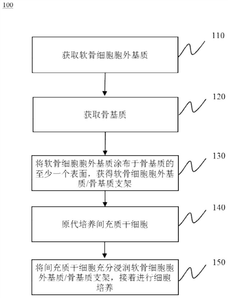
[0038] Example 1 Preparation of Chondrocyte Extracellular Matrix / Bone Matrix-Adipose Mesenchymal Stem Cells (DCECM / DBM-rADMSCs) Composite Scaffold

[0039] 【Preparation of Chondrocyte Extracellular Matrix / Bone Matrix (DCECM / DBM) Scaffold】

[0040] 1-1 Preparation of chondrocyte extracellular matrix (DCECM)

[0041] (1) Take out 100 g of fetal bovine articular cartilage one day old, and wash it with sterile distilled water for 3 times, each time for 10 minutes;

[0042] (2) Soak in 1% acetic acid for 3 times, each time for 3 hours;

[0043] (3) Hypotonic freezing and thawing in a freezer at -20°C for 8 hours;

[0044] (4) Melt ice, discard water and wash with distilled water twice;

[0045] (5) soak and sterilize in 3% hydrogen peroxide solution for 6 hours;

[0046] (6) Washing with sterile three-distilled water for 3 times, each time for 10 minutes;

[0047](7) Pulverize in a pulverizer to a homogenate with cells in millimeter, micron, and nanometer scales;

[0048] (...

Embodiment 2

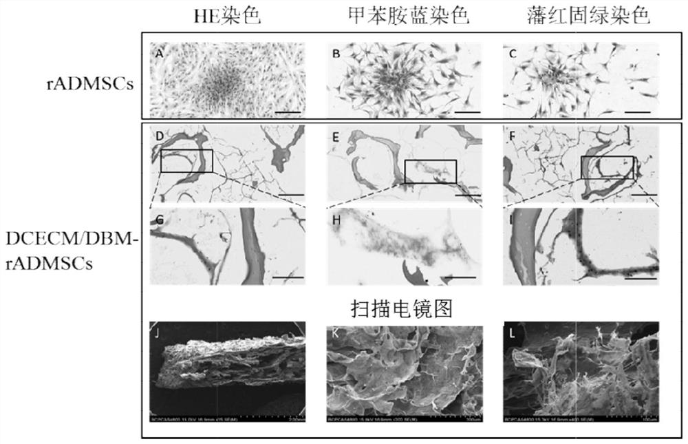
[0078] Example 2 Histopathological examination of chondrocyte extracellular matrix / bone matrix-rabbit adipose-derived mesenchymal stem cells (DCECM / DBM-rADMSCs) composite scaffold

[0079] 2-1 Prepare chondrocyte extracellular matrix / bone matrix (DCECM / DBM) scaffold in the same manner as in Example 1

[0080] 2-2 Primary culture of rabbit adipose-derived mesenchymal stem cells (rADMSCs)

[0081] (1) Sumianxin + ketamine (1:1) is anesthetized by intramuscular injection of 0.3ml / kg;

[0082] (2) Put the rabbit on the operating table, remove the hair in the operation area, disinfect the operation field with iodophor cotton ball three times, and wipe with alcohol three times;

[0083] (3) Make an incision about 3 cm long in the right lower groin of the rabbit, the depth reaches the subcutaneous tissue, bluntly separate the subcutaneous tissue, explore while separating until the subcutaneous fat is found, carefully cut out the blocky fat tissue, and pay attention to avoiding l...

Embodiment 3

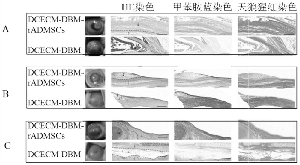
[0100] Example 3 Animal experiment of cartilage generation effect of chondrocyte extracellular matrix / bone matrix-rabbit adipose-derived mesenchymal stem cells (DCECM / DBM-rADMSCs) composite scaffold

[0101] 3-1 Prepare chondrocyte extracellular matrix / bone matrix-rabbit adipose-derived mesenchymal stem cells (DCECM / DBM-rADMSCs) composite scaffold

[0102] 3-2 Rabbit corneal chondrocyte extracellular matrix / bone matrix-rabbit adipose-derived mesenchymal stem cells (DCECM / DBM- rADMSCs) composite scaffold autografting

[0103] (1) Lu Mianxin: Midazolam = 1:1, 0.2ml / Kg intramuscular injection for anesthesia;

[0104] (2) Place the rabbit on the operating table, sterilize the right eye with iodophor, deiodine with alcohol, spread a sterile drape, place a lid speculum, and treat the eye with Benuoxi;

[0105] (3) scrape off the corneal epithelium with a corneal epidermectomy;

[0106] (4) Make a lamellar incision about 3 mm long at a distance of 2 mm from the corneal limbu...


PUM

PropertyMeasurementUnit
thicknessaaaaaaaaaa
diameteraaaaaaaaaa

Abstract

The invention provides a stent for keratoprosthesis and a preparation method thereof. The stent comprises: decalcified and decellularized cancellous bone matrix slices->dipping and sticking nanoscale extracellular matrixes (ECM) derived from the cartilage, the umbilical cord and other sources inside and outside the stent->compound autologous mesenchymal stem cells (bone mesenchymal stem cells, adipose-derived mesenchymal stem cells, etc.). After culture, the survived keratoprosthesis stent is placed between layers, on the anterior surface, on the conjunctiva or under the mucosa of the cornea, in order to be used for replacing bones, cartilages or tooth bones in an keratoprosthesis operation, and to be used as a stent/reinforcement material of the keratoprosthesis. According to the invention, the stent for the keratoprosthesis has a microenvironment which is beneficial to in-vitro and in-vivo growth of cells through adsorption and induction, can promote mesenchymal stem cells to be differentiated into cartilaginous tissues and osteogenic tissues in vivo, thus being convenient to fix the keratoprosthesis and improve the in-situ rate of the keratoprosthesis.

Description

technical field [0001] The invention relates to the technical field of artificial corneas, in particular to a bracket for artificial corneas and a preparation method thereof. Background technique [0002] Artificial cornea (Keratoprosthesis, Kpro) is an artificial device used to replace the diseased cornea to exert its optical and biological functions. The artificial cornea generally includes two parts: the lens column (optical part) and the bracket (reinforcement material). Among them, the lens column is made of a transparent material with excellent optical properties and stable physical and chemical properties, which is used to replace the turbidity that hinders the optical pathway of the eye after the lesion cornea. In order to achieve the ideal optical effect, most of the artificial corneas currently adopt the penetrating type, that is, the lens column of the artificial cornea penetrates the whole layer of the cornea, and the lens column is fixed in the center of the co...

Claims


Application Information

Patent Timeline
Patent Type & Authority Applications(China)
IPC IPC(8): A61L27/36A61L27/38
CPCA61L27/3633A61L27/3641A61L27/3687A61L27/3691A61L27/3608A61L27/3834A61L2430/16A61L2430/40
Inventor 王丽强黄一飞黄靖香程帅帅
Owner THE FIRST MEDICAL CENT CHINESE PLA GENERAL HOSPITAL
Why Eureka
Try Eureka