Novel opto-acoustic-ultrasonic bimodal transrectal endoscopic imaging probe for clinical use

An imaging probe, clinical technology, applied in ultrasound/acoustic/infrasonic image/data processing, endoscope, proctoscope, etc., can solve the lack of physical information, reduce the real-time imaging rate of two-dimensional tissue surface, small space, etc. problems, to achieve the effect of convenient and fast operation and rich organizational signal information

Pending Publication Date: 2021-07-30
TONGJI UNIV
View PDF4 Cites 0 Cited by
  • Summary
  • Abstract
  • Description
  • Claims
  • Application Information

AI Technical Summary

Problems solved by technology

[0006] However, at present, there is a lack of devices for simultaneous non-destructive endoscopic detection of prostate and other tissues through the rectum. The spy detection probe needs to be of appropriate size and should not be too large
[0007] Among the existing patents, Chinese patent CN107638168A discloses an endoscope for photoacoustic detection of intestinal tissue. The disadvantage of this device is that it can only perform photoacoustic imaging of intestinal tissue and lacks physical information of the tissue; although there are similar Chinese patent CN103385758A discloses an endoscope for ultrasonic-photoacoustic detection of the inside of blood vessels. However, the disadvantage of this device is that it belongs to destructive detection and can only collect single-point signals. To obtain two-dimensional tissue information, it needs Rotating multi-point acquisition signals and synthesizing images greatly reduces the rate of real-time imaging of two-dimensional tissue surfaces; Chinese patent CN106264604A discloses a full-scan photoacoustic dual-mode endoscopic probe, which can realize photoacoustic endoscopic inspection of intestinal tissue imaging, but cannot perform photoacoustic ultrasound dual-mode imaging on organs with a greater detection depth (penetrating the intestinal wall), such as the prostate; Chinese patent CN109199332A discloses a photoacoustic and ultrasound dual-mode internal Looking at the imaging device and method, the acoustic head is a multi-element linear array, which can realize the imaging of a larger area to be tested, realize the rapid imaging of the entire tissue to be tested, and improve the detection efficiency, but its control circuit board is located in the detection area at the front end of the probe. There is very little space for the optical module in the limited space, and it is impossible to realize the light guide of high-power hard optical fiber

Method used


Image

  • Novel opto-acoustic-ultrasonic bimodal transrectal endoscopic imaging probe for clinical use
  • Novel opto-acoustic-ultrasonic bimodal transrectal endoscopic imaging probe for clinical use
  • Novel opto-acoustic-ultrasonic bimodal transrectal endoscopic imaging probe for clinical use

Examples


Embodiment Construction

[0039] The present invention will be described in detail below in conjunction with the accompanying drawings and specific embodiments. This embodiment is carried out on the premise of the technical solution of the present invention, and detailed implementation and specific operation process are given, but the protection scope of the present invention is not limited to the following embodiments.

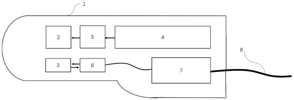
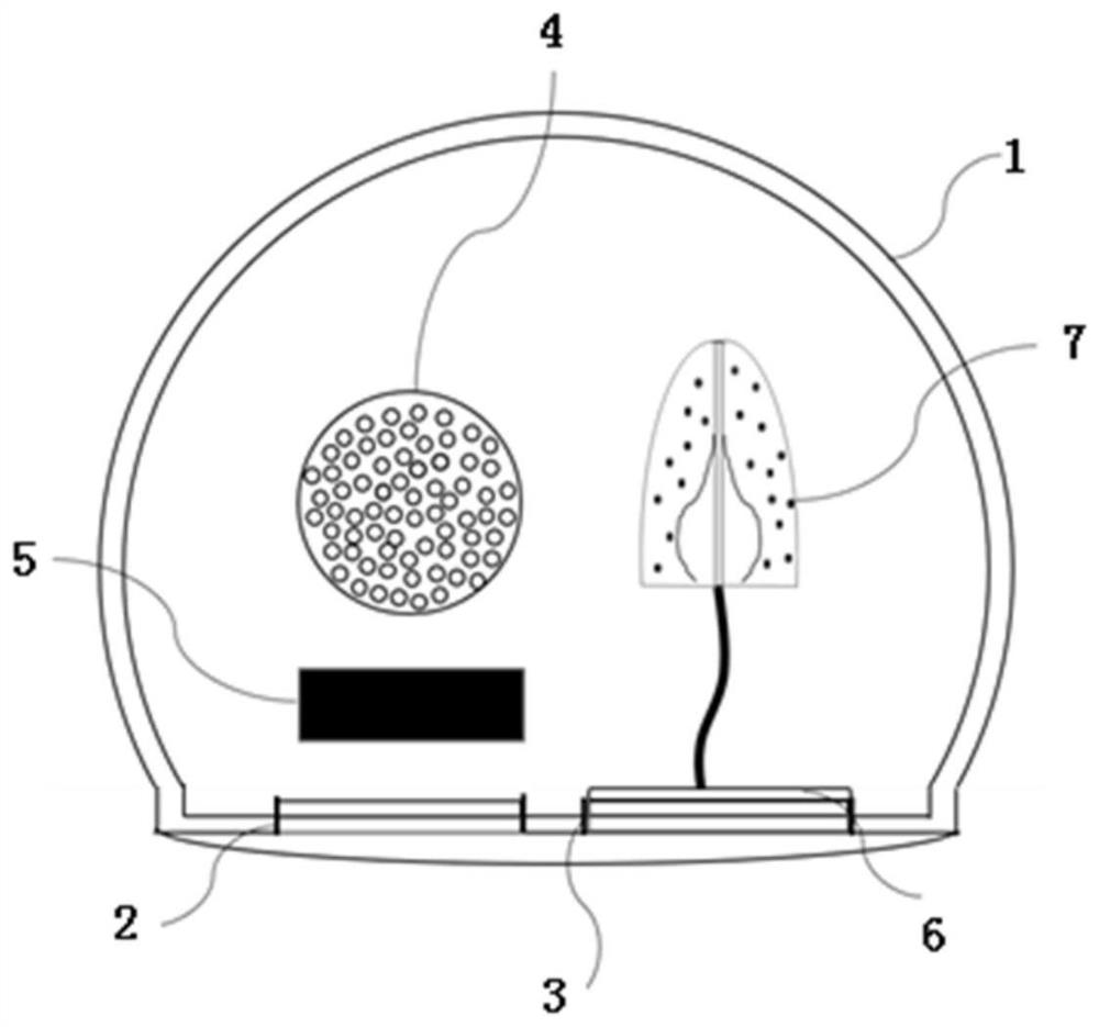
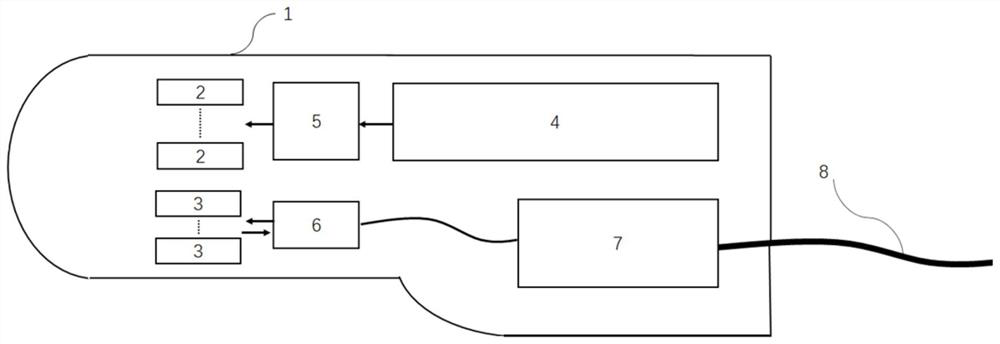
[0040] The present invention provides a novel photoacoustic ultrasound dual-mode transrectal endoscopic imaging probe for clinical use. It is difficult to detect the photoacoustic and ultrasonic dual-mode signals on the hardware and integrate it into the same endoscopic probe device when the scale is small enough. And to provide higher luminous efficiency, the invention realizes synchronous and co-located collection of ultrasonic and photoacoustic signals by relocating the control circuit at the handle end of the probe and combining the optical module and the acoustic module.

[0041]...


PUM


Abstract

The invention relates to a novel clinical opto-acoustic ultrasonic bimodal transrectal endoscopic imaging probe, which comprises a probe shell that comprises a front-end detection part and a rear-end handheld part which are integrally formed, wherein the side surface of the front-end detection part is provided with an optical signal transmitting window and an acoustic signal transmitting and receiving window which are arranged in parallel; an optical module, which is arranged in the probe shell and is used for emitting uniformly-shaped light beams and irradiating a to-be-imaged tissue area through the light beam emitting window to generate a photoacoustic signal; and an ultrasonic module, which, along with the optical module, is arranged in the probe shell together and is used for transmitting an ultrasonic signal to irradiate the to-be-imaged tissue area, which is as same as the optical signal transmitting window through the acoustic signal transmitting and receiving window, and receiving the photoacoustic signal and the reflected ultrasonic signal at the same time. Compared with the prior art, the device has the advantages of meeting the size requirement of ergonomics, noninvasive detection, small size, photoacoustic and ultrasonic dual-mode detection and the like.

Description

technical field [0001] The invention relates to the fields of endoscope and non-destructive testing, in particular to a novel photoacoustic ultrasonic dual-mode transrectal endoscopic imaging probe for clinical use. Background technique [0002] Ultrasound imaging for medical diagnosis is widely used in medical diagnosis due to its advantages of non-ionizing radiation, high-resolution imaging of anatomical structures, and low cost. However, ultrasound imaging is generally sensitive to tissue structures and blood flow with differences in acoustic impedance, and has no specificity to other physical and chemical properties of tissues, so the diagnostic function is limited. In many medical clinical diagnoses, ultrasound imaging needs to be combined with MRI, CT or X-ray imaging. Combined with other radioactive diagnostic methods such as light to provide doctors with more specific lesion information. [0003] Transrectal endoscopic ultrasound imaging probes have been widely used...

Claims


Application Information

Patent Timeline
Patent Type & Authority Applications(China)
IPC IPC(8): A61B5/00A61B8/08A61B8/12A61B1/31A61B1/07
CPCA61B1/07A61B1/31A61B5/0035A61B5/004A61B5/0095A61B8/0833A61B8/12A61B8/4444A61B8/4488A61B8/5261
Inventor 程茜解维娅张浩南李佳琰陈盈娜张梦娇高雅吴诗颖王一凡
Owner TONGJI UNIV
Why Eureka
Try Eureka