Method for detecting biological activity of granulocyte colony stimulating factors

A technology of colony stimulating factor and biological activity, applied in the field of biomedicine, can solve the problems of experimental failure, long operation time and high operation requirements, and achieve the effect of shortening the experimental operation time, shortening the operation time and improving the detection throughput.

Pending Publication Date: 2021-11-26
江苏奥赛康生物医药有限公司
View PDF0 Cites 0 Cited by
  • Summary
  • Abstract
  • Description
  • Claims
  • Application Information

AI Technical Summary

Problems solved by technology

At present, the biological activity of G-CSF, PEG-GCSF, etc. is detected by NFS60 cell proliferation assay. This method has the following disadvantages: the operation process is relatively complicated, such as the need to add stimulatory factors in advance to co-culture cells; the cell color development process uses CCK8, 10 μL / well, the solution volume is small, the operation requirements are high, and it is easy to make mistakes, which leads to the failure of the experiment; the operation time is long, and the whole experiment process takes 3 days to complete

Method used


Image

  • Method for detecting biological activity of granulocyte colony stimulating factors
  • Method for detecting biological activity of granulocyte colony stimulating factors
  • Method for detecting biological activity of granulocyte colony stimulating factors

Examples


Embodiment 1

[0045] Experimental reagents:

[0046] Complete medium: 10% FBS (mass fraction) + DMEM + 300 μg / mL Hygromycin B + 700 μg / mL Geneticin

[0047] Working medium: 1% FBS (mass fraction) + DMEM

[0048] Plasmid pGL4.47-STAT3-Luc-HygromycinB: purchased from Promega, Cat. No. E4047

[0049] Chromogenic agent Bright glo: purchased from Promega company

[0050] 0.25% trypsin: purchased from Gibco company, unless otherwise specified in the present invention, trypsin solution refers to the weight to volume ratio, specifically refers to 0.25g trypsin in 100mL solution

[0051] Experimental sample:

[0052] Standard product: rhG-CSF from China National Institutes for Food and Drug Control, batch number 98 / 01

[0053] The test product: G-CSF or PEG-GCSF is from Jiangsu Aosaikang Pharmaceutical Co., Ltd. Experimental equipment:

[0054] CO 2Incubator: Thermo, HERA cell 150i

[0055] Cell counter: CounterStar, IC1000

[0056] Microplate reader: Biotck, synergy H1, Gen5 software system...

Embodiment 2

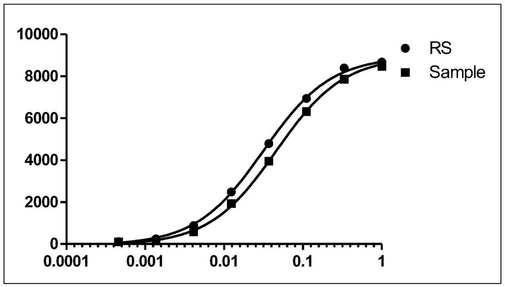
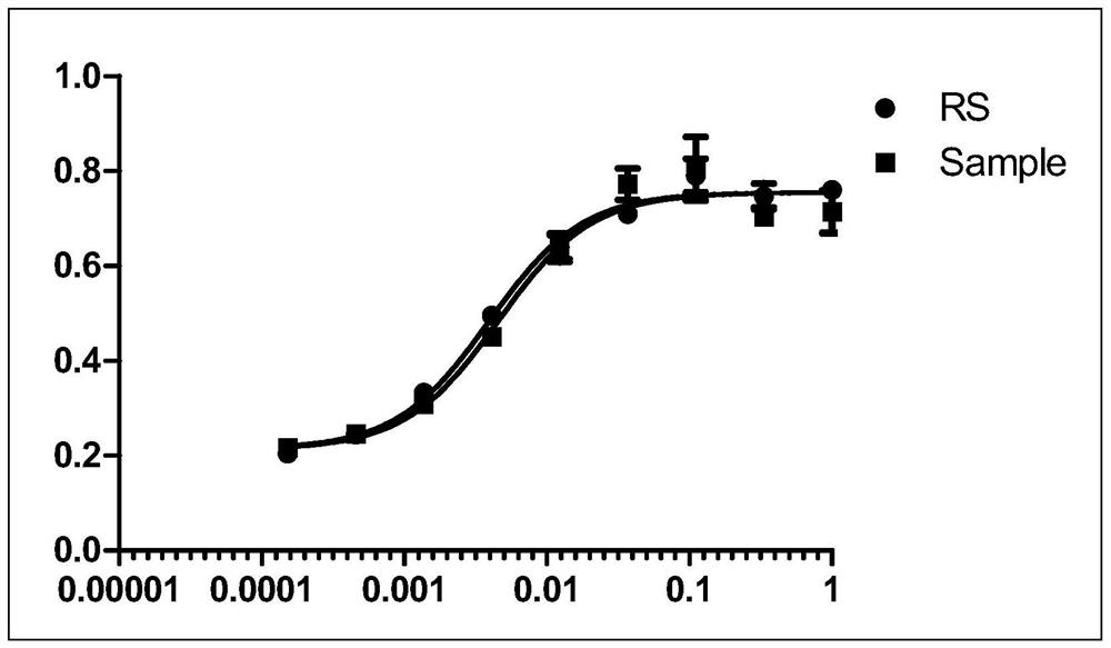
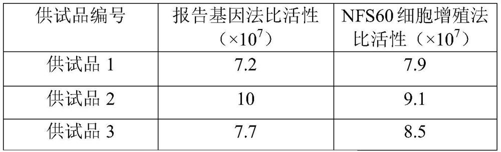
[0079] The method of Example 1 and the NFS60 cell proliferation method were used to detect three groups of test products (G-CSF) respectively, and the accuracy of the method of the present invention was verified.

[0080] Experimental reagents:

[0081] Basal medium: 90% RPMI-1640 medium (mass fraction) + 10% FBS (mass fraction) + 55mM2-Mercaptoethanol

[0082] Complete medium: basal medium + final concentration 20ng / mL rhG-CSF

[0083] RPMI-1640 medium and 2-Mercaptoethanol were purchased from Gibco

[0084] M-NFS-60 cells: from ATCC

[0085] Experimental sample:

[0086] Standard product: rhG-CSF from China National Institutes for Food and Drug Control, batch number 98 / 01

[0087] The specific steps of the NFS60 cell proliferation method are as follows:

[0088] 1. Cell Culture and Assay Plate Preparation

[0089] M-NFS-60 cell culture:

[0090] 1) Pre-warmed culture medium

[0091] 2) Take one tube of frozen M-NFS-60 cells, dissolve it quickly at 37°C, and add it to...


PUM

PropertyMeasurementUnit
correlation coefficientaaaaaaaaaa

Abstract

The invention provides a method for detecting biological activity of granulocyte colony stimulating factors. The method comprises the following three steps: constructing a cell line, preparing a standard substance and a test sample, and determining. The detection method is more convenient to operate, cells do not need to be specially treated, the operation time is shortened, and the experiment operation time is shortened by 1-2 days; meanwhile, according to the method, the detection flux is improved, the sample is diluted by 6-12 concentration points, the sample can be vertically arranged in a 96-well plate, and the sample detection number of each cell plate is increased.

Description

technical field [0001] The invention relates to the technical field of biomedicine, in particular to a method for detecting the biological activity of granulocyte colony-stimulating factor. Background technique [0002] Human granulocyte colony stimulating factor (hG-CSF) is a hematopoietic growth factor that regulates the maturation and growth of granulocytes (especially neutrophils) in mammals. It promotes the release of mature neutrophils into the peripheral blood by stimulating the differentiation and maturation of granulocyte-macrophage colony-forming units (CFU-GM) to granulocyte-colony-forming units (CFU-G), thereby promoting neutrophil growth. It is clinically used to treat neutropenia caused by various reasons, which has great economic and social significance. [0003] In 1985, Welte.K successfully purified and refined human granulocyte colony-stimulating factor (hG-CSF) from the culture supernatant of human bladder cancer cell line 5637, and then Welte.K cooperat...

Claims


Application Information

Patent Timeline
Patent Type & Authority Applications(China)
IPC IPC(8): G01N21/64
CPCG01N21/6486
Inventor 赵俊郑海亮方雅王东方袁云霞文勇
Owner 江苏奥赛康生物医药有限公司
Why Eureka
Try Eureka