
Endoplasmic reticulum membrane fusion liposome as well as preparation method and application thereof

An endoplasmic reticulum membrane and liposome technology, applied in the biological field, can solve the problems of complex chemical reaction, high exploration cost, unfavorable clinical transformation, etc., and achieve the effect of reducing Zeta potential and facilitating industrial transformation.

Active Publication Date: 2021-12-24
SHANDONG UNIV
View PDF1 Cites 0 Cited by
  • Summary
  • Abstract
  • Description
  • Claims
  • Application Information

AI Technical Summary

Problems solved by technology

However, the discovery and design of these targeting molecules depend on the comprehensive analysis of target proteins located on the endoplasmic reticulum, and the cost of exploration is too high; well-designed synthetic materials are not conducive to clinical practice due to their uncertain biocompatibility and complex chemical reactions. convert

Method used


Image

  • Endoplasmic reticulum membrane fusion liposome as well as preparation method and application thereof
  • Endoplasmic reticulum membrane fusion liposome as well as preparation method and application thereof
  • Endoplasmic reticulum membrane fusion liposome as well as preparation method and application thereof

Examples


Embodiment 1

[0039] Example 1: Extraction of endoplasmic reticulum membrane in B16F10 cells

[0040] Prepare 10 flasks of B16F10 cells in T75 flasks (count 5*10 7 ), after washing twice with PBS buffer, each bottle was digested with 3 mL of trypsin ethylenediaminetetraacetic acid hydrochloride for 2 min, and the digestion was terminated by adding DMEM complete medium, and the cell digestion solution was collected and centrifuged at 300 g for 5 min. Discard the supernatant and save the cell pellet. Add 2 mL of pre-cooled grinding buffer (sigma) and protease inhibitor (Beiyuntian), transfer to a pre-cooled DOUNCE homogenizer, and homogenize on ice for 50 times until more than 80% of the cells are lysed under the microscope. Transfer the homogenate to a centrifuge tube, centrifuge at 1000g at 4°C to remove nuclei and cell debris, and collect the supernatant. Then the supernatant was centrifuged at 4° C. and 12000 g for 30 min, and the supernatant was collected. Use an ultracentrifuge and s...

Embodiment 2

[0042] Example 2: Preparation of doxorubicin (DOX)-loaded liposomes by ammonium sulfate gradient method and preparation of EM / DOX Lip by ultrasonic mixing method

[0043] First, dissolve DOPC, DOPE, and CHEMS in 3ml chloroform at a ratio of 1:1:0.5, place in a rotary evaporator and evaporate under reduced pressure. The temperature of the water bath is 25°C and the speed is 70rpm / min. After forming a transparent and uniform lipid film on the inner wall, vacuum dry overnight to completely remove residual solvent. Add 5 mL of ammonium sulfate solution (250 mmol, pH = 5.5) preheated to 40 ° C and sonicate in a water bath to obtain a liposome solution with light blue opalescence. Then the obtained solution was put into a treated dialysis bag and dialyzed for 48 hours to remove free ammonium sulfate, and diluted to 1 mg / mL liposome solution.

[0044] Then add 0.1 mg of free doxorubicin solution per milliliter to the resulting solution, heat it in a water bath to 40°C and rotate and...

Embodiment 3

[0049] Example 3: Extraction of DC2.4 Cell Endoplasmic Reticulum Membrane

[0050] Prepare 8 bottles of T75 culture flasks of cells (count 4*10 7), washed twice with PBS buffer, digested with trypsin ethylenediaminetetraacetic acid hydrochloride for 3 minutes, added complete medium to terminate the digestion, and collected the cell digestion solution and centrifuged at 200g for 10 minutes. Discard the supernatant and save the cell pellet. Add 1mL of pre-cooled grinding buffer (sigma) and protease inhibitor (Beiyuntian), transfer to a pre-cooled DOUNCE homogenizer, and homogenize on ice for 20 times until most of the cells are lysed under a microscope. Transfer the homogenate to a centrifuge tube, centrifuge at 1000g at 4°C to remove nuclei and cell debris, and collect the supernatant. Then the supernatant was centrifuged at 12000 g for 15 min at 4° C., and the supernatant was collected. Use an ultracentrifuge and supporting tubes to centrifuge again at 4°C and 150,000g for ...


PUM

PropertyMeasurementUnit
particle diameteraaaaaaaaaa

Abstract

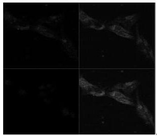
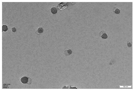
The invention relates to an endoplasmic reticulum membrane fusion liposome as well as a preparation method and application thereof, and belongs to the technical field of biology. The method comprises the following steps: (1) extraction of endoplasmic reticulum membrane; (2) preparation and characterization of lipidosome; and (3) preparation and characterization of the endoplasmic reticulum membrane fusion liposome. The liposome prepared by using a film dispersion method has a particle size controlled to be 103.03 + / -7.99 nm, a narrow particle size distribution, PDI of 0.149 + / -0.02, and a zeta potential of 41.8 + / -2.71 mV. After the liposome is fused with the endoplasmic reticulum membrane, the final endoplasmic reticulum membrane fused liposome (EM / Lip) has a particle size controlled to be 124.27 + / -4.21 nm, PDI of 0.19 + / -0.03, and a zeta potential of 14.27 + / -2.50 mV. Most of the EM / Lip is positioned in the endoplasmic reticulum after entering the cells, and little EM / Lip enters the lysosome, so that degradation of the drug in the lysosome is avoided, and a targeting function of the endoplasmic reticulum is realized. The endoplasmic reticulum membrane fusion liposome obtained by the invention can be applied to a endoplasmic reticulum targeting function of immune cells and tumor cells, and the laser confocal three-dimensional image technology is also well applied to the research on the distribution of subcellular organelle level nano preparations.

Description

technical field [0001] The invention relates to an endoplasmic reticulum membrane fusion liposome and a preparation method and application thereof, belonging to the field of biotechnology. Background technique [0002] With the development of precision medicine, the development of appropriate drugs and drug delivery systems based on key biological macromolecules, such as genes and proteins, that occur in the disease itself has become the latest research hotspot. And most of the key disease-related biomolecules are located intracellularly. Only precise targeting can achieve precise life regulation. An attractive strategy to improve the therapeutic index of drugs is: not only to carry therapeutic agents to target tissues and cells, but also to control the precise targeting of drugs to subcellular structures. Realize precise drug distribution and release of intracellular targets. However, most studies on drug delivery systems to date have struggled to overcome extracellular b...

Claims


Application Information

Patent Timeline
Patent Type & Authority Applications(China)
IPC IPC(8): A61K9/127A61K45/00A61K31/704A61K31/7088A61K47/46A61P35/00C12N5/071C12N5/09
CPCA61K9/1271A61K9/1277A61K45/00A61K31/704A61K31/7088A61K47/46A61P35/00C12N5/0693C12N5/0626C12N2509/00C12N2509/10
Inventor 林贵梅傅相蕾邱胜男
Owner SHANDONG UNIV
Learn More