Construction method of self-protection DNA enzyme walker and application of self-protection DNA enzyme walker to living cell miRNA detection

A DNA enzyme and construction method technology, applied in the field of construction of self-protected DNA enzyme walker, to achieve high specificity, good stability against degradation, and increase the effect of cleavage rate

Pending Publication Date: 2022-01-18
FUZHOU UNIV
View PDF11 Cites 2 Cited by
  • Summary
  • Abstract
  • Description
  • Claims
  • Application Information

AI Technical Summary

Problems solved by technology

Therefore, developing a DNase walker for long-term intracellular miRNA imaging remains a formidable challenge for practical application.

Method used


Image

  • Construction method of self-protection DNA enzyme walker and application of self-protection DNA enzyme walker to living cell miRNA detection
  • Construction method of self-protection DNA enzyme walker and application of self-protection DNA enzyme walker to living cell miRNA detection
  • Construction method of self-protection DNA enzyme walker and application of self-protection DNA enzyme walker to living cell miRNA detection

Examples


Embodiment 1

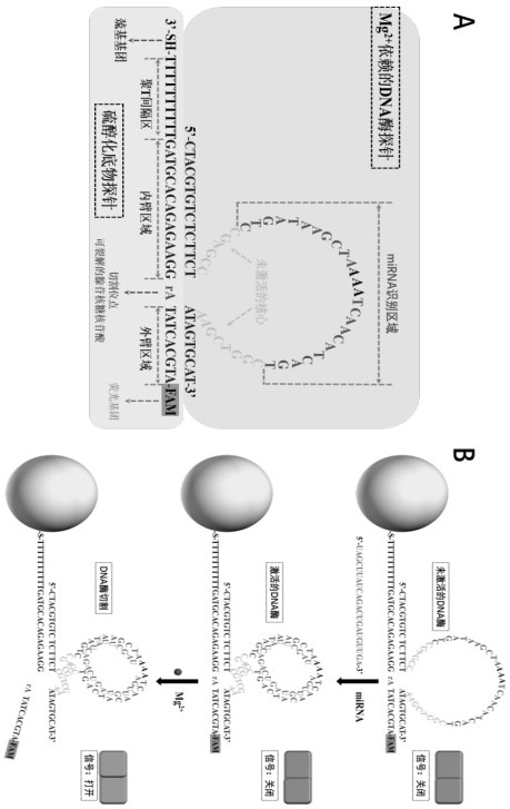
[0039] Example 1 A method for preparing a self-protected DNase walker, comprising the following steps:

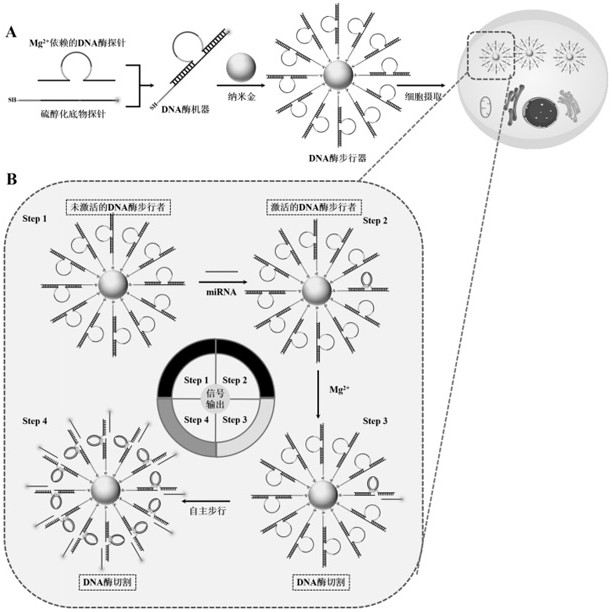
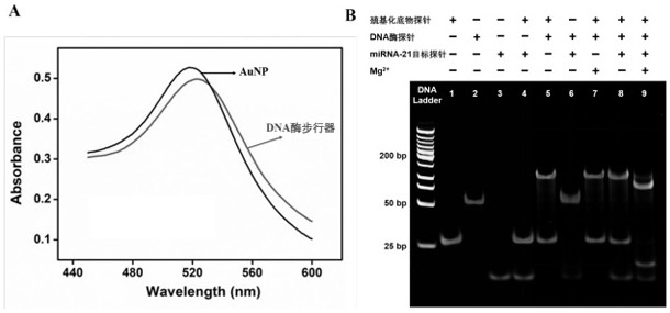
[0040] (1) First, gold nanoparticles (AuNPs) with a diameter of 13 nm were prepared by the sodium citrate reduction method. Next, in 19.5 µL of 500 mM acetate buffer (Tris-Ac buffer pH 5.2), 0.5 µL of 10 mM tris(2-carboxyethyl)phosphine was mixed with 30 µL of 100 µM thiolated substrate chain probe, and the Incubate for 1 hour at room temperature to reduce sulfhydryl groups.

[0041] (2) Add 3 µL of 100 µM Mg to the reduced thiolated substrate chain solution 2+DNase-dependent probes, followed by incubation for 3 hours at 37 °C for complete hybridization. The molar ratio of the two nucleic acids is 1:10 (substrate probe: DNase probe). Then, the resulting mixture was added to a clean glass vial containing 1 mL of AuNP solution (10 nM), and the glass vial was incubated with shaking in the dark (200 rpm) for 16 h. After the incubation time has elapsed, add 10.3 µL of 500 mM...

Embodiment 2

[0047] Example 2 Feasibility analysis of a self-protected DNase walker for in vitro detection of miRNA

[0048] Using the self-protected DNase walker constructed by the method in Example 1, the feasibility analysis was performed with the miRNA-21 target probe.

[0049] The miRNA-21 target probe sequence used is: 5'-TAGCTTATCAGACTGATGTTGA-3'.

[0050] The specific steps of feasibility analysis are as follows:

[0051] First, add Mg to 18 μL Tris-Ac Buffer solution with pH 5.2 2+ Dependent DNase probe (0.1 μM) and thiolated substrate probe (1 μM), then incubated at 37 °C for 3 h to allow Mg 2+ The dependent DNase probe fully hybridizes to the thiolated substrate probe. Next, add 1 μL of 2 μM miRNA-21 target probe to activate the DNase machinery. After incubation at 37 °C for 30 min, add 1 μL of 200 mM Mg 2+ to initiate DNase-catalyzed cleavage. The resulting solution was incubated at 37°C for 8 hours. Other samples, including blanks and controls, were prepared with corres...

Embodiment 3

[0054] Example 3 Analysis of the detection ability of the self-protected DNase walker to the target DNA probe

[0055] A self-protected DNase walker was constructed using the method in Example 1, by adding different concentrations of target DNA probes, and then using a fluorescence spectrometer to test its kinetics. Specific steps are as follows:

[0056] Under the condition of 37 ̊C, the time-varying curves of fluorescence signals of different concentrations of miRNA-21 target probe samples and blanks were monitored, and the scanning time was 1 h. The excitation wavelength was 488 nm and the emission wavelength was 520 nm. Specifically, a final concentration of 2 nM of DNase walker and different concentrations of miRNA-21 target probe (final concentrations of 0, 100 pM, 600 pM, 1 nM, 4 nM, 8 nM, 10 nM, 12 nM and 15 nM), and adjust the volume to 190 μL with Tris-Ac buffer (pH = 8.2). After the mixed solution was incubated at 37 ̊C for 30 minutes, 10 μL of Mg 2+ solution (fi...


PUM

PropertyMeasurementUnit
diameteraaaaaaaaaa

Abstract

The invention provides a construction method of a self-protection DNA enzyme walker and an application of the self-protection DNA enzyme walker to living cell miRNA detection, and belongs to the technical field of nano materials. The self-protection DNA enzyme walker comprises nanogold, a thiolated substrate probe and an Mg <2+> dependent DNA enzyme probe. The self-protection DNA enzyme walker can drive the self-protection DNA enzyme walker to gradually move on a gold nanoparticle track through specific substrate probe cutting so as to realize high-sensitivity intracellular imaging of an miRNA biomarker. The self-protection DNA enzyme walker can accurately distinguish diseased cells from healthy cells, due to the designability of DNA, the DNA enzyme walker can be expanded to image other miRNAs only by changing a target binding domain, and the self-protection DNA enzyme walker is a promising cancer diagnosis and prognosis tool.

Description

technical field [0001] The invention belongs to the technical field of nanomaterials, and in particular relates to a construction method of a self-protected DNA enzyme walker and its application in the detection of living cell miRNA. Background technique [0002] MicroRNA (miRNA, about 22 nucleotides) is a class of RNA molecules found in animals, plants and some viruses. It has endogenous, non-coding and regulatory characteristics, and plays an important role in RNA silencing and gene regulation. Role. A growing body of research evidence has shown that abnormal expression of miRNAs is associated with the occurrence of various cancers and many other pathological conditions. For example, miRNA-21 is closely related to the proliferation, apoptosis, invasion and migration of malignant tumors, and is often overexpressed in chronic lymphocytic leukemia, cervical cancer, lung cancer, breast cancer and prostate cancer. Therefore, miRNAs become one of the most promising targets for...

Claims


Application Information

Patent Timeline
Patent Type & Authority Applications(China)
IPC IPC(8): C12Q1/6825C12N9/22C12N15/11
CPCC12Q1/6825C12N9/22
Inventor 吴再生王伟军高艳莎王文青
Owner FUZHOU UNIV
Why Eureka
Try Eureka