
Method for promoting transdifferentiation of pAdM3C infected rat pancreatic duct cells

A pancreatic duct and transdifferentiation technology, applied in the direction of pancreatic cells, cell dissociation methods, biochemical equipment and methods, etc., can solve the problems of difficult culture, complicated identification, low cell acquisition rate, etc., and achieve the goal of increasing the number and purity of cells Effect

Pending Publication Date: 2022-03-18
FUJIAN ACAD OF MEDICAL SCI
View PDF9 Cites 0 Cited by
  • Summary
  • Abstract
  • Description
  • Claims
  • Application Information

AI Technical Summary

Problems solved by technology

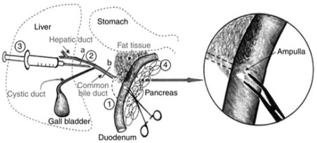
Pancreatic ductal cells are difficult to obtain, difficult to cultivate, and complicated to identify, so there are few research groups. At present, the primary cells obtained by direct digestion with digestive enzymes are more commonly used. On the one hand, the cell acquisition rate is too low, on the other hand, the cell purity Not enough, there are many fibroblasts, acinar cells, islet cells, etc.
At present, there is no report on the transdifferentiation of pancreatic ductal cells using adenovirus

Method used


Image

  • Method for promoting transdifferentiation of pAdM3C infected rat pancreatic duct cells
  • Method for promoting transdifferentiation of pAdM3C infected rat pancreatic duct cells
  • Method for promoting transdifferentiation of pAdM3C infected rat pancreatic duct cells

Examples


Embodiment 1

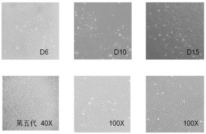
[0051] Example 1 Separation and purification of rat pancreatic ductal cells

[0052] 1. Preparation 2-3 days before the experiment

[0053] 1 Islet Isolation Reagent:

[0054] 1) Hanks solution: (Gibco, 14025) 500 ml / bottle, or prepare Ca-free 2+ , Mg 2+ Hanks solution, stored at 4°C after sterilization.

[0055] 2) 25% BSA (Bovine Serum Albumin): 1ml aliquot and store at -20°C.

[0056] 3) 12.5mg / ml collagenase storage solution: (collagenase type IX, Sigma, C-7657-1G) 1ml aliquots, store at -20°C.

[0057] 4) 0.67g / l DTZ stain stock solution: aliquot 50μl and store at -20°C.

[0058]5) Citrate buffer: citric acid (Citric acid, Sigma, C2404-100G), filter sterilized, aliquot 1ml, store at -20°C.

[0059]

[0060] Pancreatic ductal cell isolation reagents:

[0061] 1) 1×PBS-CMF: calcium and magnesium free, cell grade (that is, PBS in this protocol) (about 200ml / rat)

[0062] 2) 0.025% trypsin: 0.25% trypsin-EDTA, diluted 10 times with PBS.

[0063]

[0064] 3) Med...

Embodiment 2

[0123] Example 2 Transdifferentiation induction using adenovirus (pAd-M3C)

[0124] Experimental steps:

[0125] 1 plate: Inoculate a 12-well plate, 2.5×10 per well 5 The pancreatic ductal cells prepared in Example 1.

[0126] 2 After 24 h, the culture medium was removed, and 500 μl of fresh medium c was added to each well.

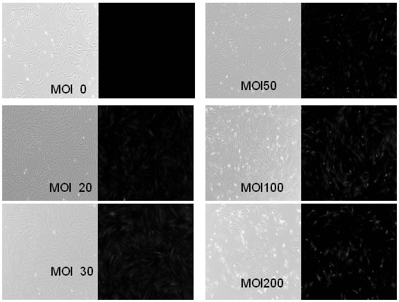
[0127] 3 Convert MOI to 20, 30, 50, 100, and 200 μl corresponding volumes of adenovirus (pAd-M3C purchased from Addgene) virus supernatant volume, respectively added in the following table.

[0128]

[0129] 4 Shake the culture plate continuously and slowly, and incubate for 3 h.

[0130] 5 Add 1.5 ml of fresh medium c and culture for 72 h.

[0131] image 3 It is to observe the situation of pancreatic ductal cells after infection under a microscope, in which the red fluorescence represents the cells that are infected and express the reporter gene. It can be seen from the figure that the pancreatic ductal cells were infected with adenovirus (pAd...


PUM


Abstract

The invention provides a method for promoting transdifferentiation of pancreatic duct cells of rats infected with pAd-M3C. Islet cell clusters can be removed as many as possible through density gradient centrifugal separation of islet by using a Biooll separating medium, cells are preliminarily separated and purified, and tube bottom cells without the islet cell clusters are taken, so that the influence of islet cells is reduced; the method comprises the following steps of: screening cells at the bottom of a catheter through a 40mu m small filter, further removing large islet cells, replacing the culture media a, b and c in different culture time periods, and promoting the growth of the catheter cells according to the proportion and action of nicotinamide, KGF and EGF in the c, so that the number and purity of the obtained cells are improved; the pancreatic duct cells are infected by the adenovirus (pAd-M3C), and the sample adding volume is verified by an infection experiment, so that the infection rate of the pancreatic duct cells is up to 80% or above, and the subsequent transdifferentiation experiment is facilitated.

Description

technical field [0001] The invention belongs to the technical field of molecular biology cell immunity. It specifically relates to a method for infecting rat pancreatic duct cells with pAdM3C to promote transdifferentiation. Background technique [0002] Separation and purification of rat pancreatic ductal cells. At present, domestic researchers mostly use digestive enzymes to directly digest and separate pancreatic tissue. By using a medium that promotes the growth of epithelial cells to screen and cultivate primary cells, the surviving cells are considered to contain Cell populations of pancreatic ductal cells. Pancreatic ductal cells are difficult to obtain, difficult to cultivate, and complicated to identify, so there are few research groups. At present, the primary cells obtained by direct digestion with digestive enzymes are more commonly used. On the one hand, the cell acquisition rate is too low, on the other hand, the cell purity Not enough, there are many fibrobl...

Claims


Application Information

Patent Timeline
IPC IPC(8): C12N5/071C12N15/861C12N15/65
CPCC12N5/0676C12N15/86C12N15/65C12N2501/734C12N2501/11C12N2501/117C12N2500/38C12N2500/34C12N2509/00C12N2710/10343C12N2800/107
Inventor 王坤韩俊永陈金烟金静君薛士杰
Owner FUJIAN ACAD OF MEDICAL SCI
Learn More