
Temporomandibular joint motion reconstruction method based on multi-modal information fusion

A temporomandibular joint and multi-modal technology, applied in the field of temporomandibular joint motion reconstruction based on multi-modal information fusion, can solve the problem of inability to directly record and observe internal structure motion, inability to intuitively reflect the real-time state of functional motion, lack of temporal Problems such as mandibular joint disc motion reconstruction, to achieve the effect of reducing image acquisition costs, reducing equipment interference, and wide application range

Pending Publication Date: 2022-07-05
天津市口腔医院(天津市整形外科医院南开大学口腔医院)
View PDF0 Cites 3 Cited by
  • Summary
  • Abstract
  • Description
  • Claims
  • Application Information

AI Technical Summary

Problems solved by technology

At present, radiological imaging technology can only obtain high-quality images by collecting static image data. Dynamic images have low resolution and are only two-dimensional imaging. Any single imaging technology cannot obtain its complete structure and disease information, and cannot intuitively reflect its Real-time status of functional movement
Mandibular movement trajectory tracing technology relies on mechanical tracing, soft tissue admissibility, in-situ measurement, etc., resulting in insufficient measurement accuracy, and cannot directly record and observe the movement of internal structures. Trajectory curves to infer the coordinated motion state of the temporomandibular joint and other soft tissues
Some researchers at home and abroad use binocular vision combined with CT technology to only display the movement of temporomandibular joint bone tissue, but lack important temporomandibular joint disk motion reconstruction, which is far from enough for temporomandibular joint research
At present, there is no system or product that can completely reconstruct the movement process of the temporomandibular joint. Dentists urgently need to intuitively understand the real-time functional status of the temporomandibular joint. This has become one of the urgent problems to be solved in the current research on temporomandibular joint diseases.

Method used


Image

  • Temporomandibular joint motion reconstruction method based on multi-modal information fusion
  • Temporomandibular joint motion reconstruction method based on multi-modal information fusion
  • Temporomandibular joint motion reconstruction method based on multi-modal information fusion

Examples


preparation example Construction

[0041] The preparation steps of the dental arch splint and the handle are as follows: making a plaster model of the lower dentition of the test-wearer, making a mandibular dentition pressure film retainer, grinding out the occlusal contact area with the maxillary dentition, obtaining the dental arch splint, The lower central incisor is attached to the handle on the labial side to facilitate holding and fixing the marker holder;

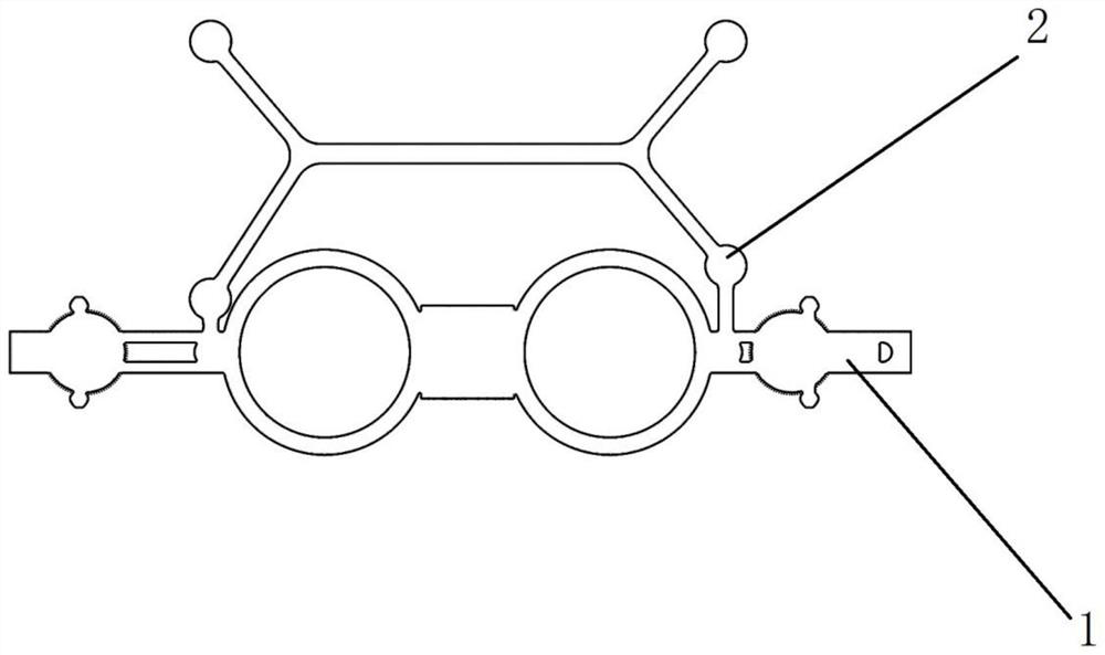
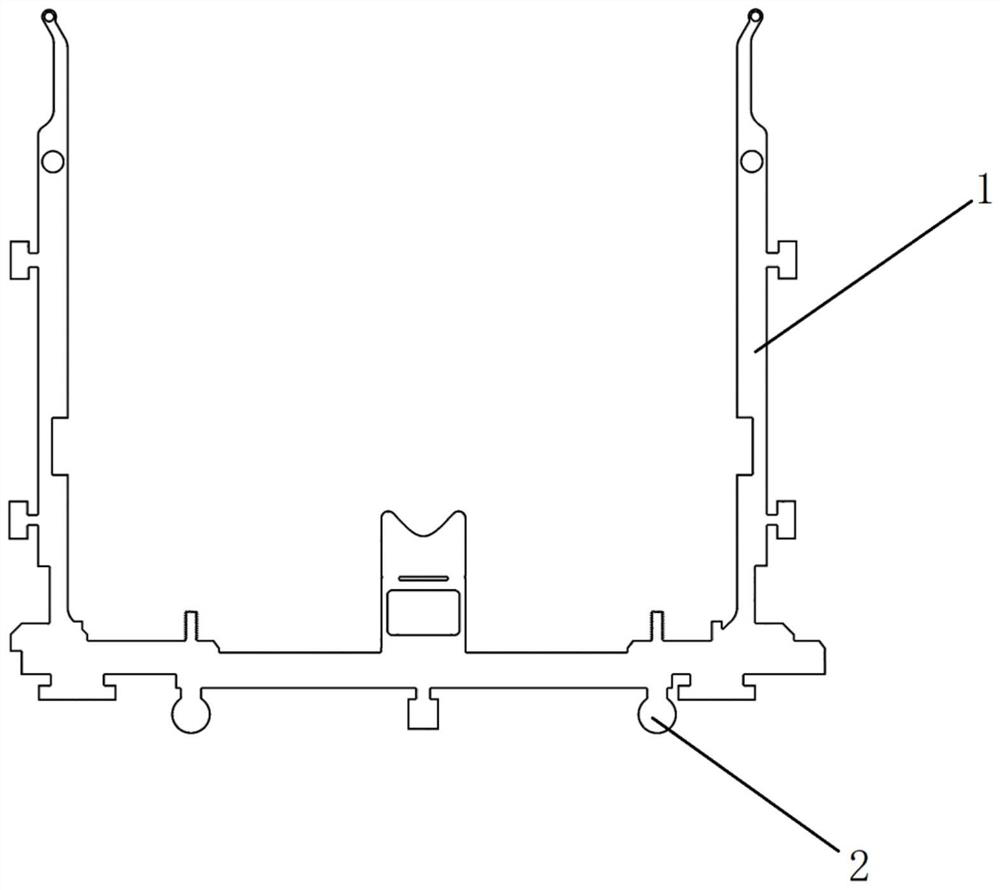
[0042] Step 13, as attached Figure 8As shown in the figure, the upper and lower jaw marking device with reflective marking balls is worn for the try-wearer: first, the upper and lower jaw marking device with four passive reflective marking balls 1 2 is worn, and the nose pads are made of silicone rubber impression material and placed on the trial fitting Adjust the distance between the left and right fixing brackets between the nasion point of the patient and the two mirror rings of the maxillary marking device to make it close to the left and right ...


PUM


Abstract

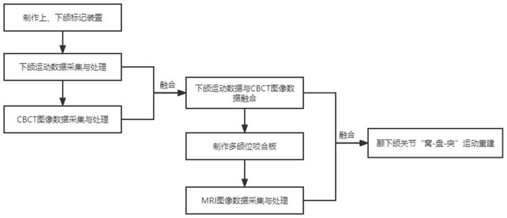
The invention provides a temporal-mandibular joint motion reconstruction method based on multi-modal information fusion. The temporal-mandibular joint motion reconstruction method is characterized by comprising the following steps: making an upper and lower jaw marking device; acquiring and processing mandibular movement data; acquiring and processing CBCT data images; fusing the mandibular movement data and the CBCT image data; a multi-jaw-position biteplate is manufactured; acquiring and processing an MRI data image; the mandibular movement data, CBCT image data and MRI image data are registered and fused, and mandibular movement is visually reproduced in a 3D model and image mode. According to the method, the ternary relative motion relation and the space posture of the temporomandibular glenoid fossa, the joint disc and the condyle process are accurately recorded, the real functional motion state of the temporomandibular joint is revealed in a visual and quantitative mode, and the method can be used for clinical research of the temporomandibular joint, serves as an auxiliary means for diagnosis, treatment and prognosis evaluation of temporomandibular joint diseases and has a good application prospect. Clinical operation is easy and convenient, the difficulty that a clinician judges the position of the joint disc can be lowered, and the accuracy of diagnosis and treatment of temporomandibular joint diseases is improved.

Description

technical field [0001] The invention relates to the field of temporomandibular joint motion reconstruction, in particular to a temporomandibular joint motion reconstruction method based on multimodal information fusion. Background technique [0002] In recent years, multi-modal information fusion technology has received extensive attention in the medical field. Among them, medical image fusion can combine different imaging data, make full use of the advantages of various medical information, and simultaneously display anatomical structure and functional status in one image. It provides more comprehensive physiological or pathological information in an intuitive form, and provides technical means for accurate diagnosis and treatment of oral-related diseases. [0003] At present, the commonly used auxiliary examination techniques for the temporomandibular joint are radiography and mandibular trajectory tracing. At this stage, radiological imaging technology can obtain high-qu...

Claims


Application Information

Patent Timeline
Patent Type & Authority Applications(China)
IPC IPC(8): G06T7/30G06T17/00G06T7/70G06T7/20G06T5/50G16H30/20G16H50/20
CPCG06T7/30G06T17/00G06T7/20G06T7/70G16H50/20G16H30/20G06T5/50G06T2207/10088G06T2207/10016G06T2207/30008G06T2207/20221
Inventor 张淋坤王鸿鹏张璐申林张春香
Owner 天津市口腔医院(天津市整形外科医院南开大学口腔医院)
Learn More