Avian pluripotent embryonic germ cell line
A cell line and germ cell technology, applied in the field of avian pluripotent embryonic germ cells, can solve the problems of no instruction or expected continuous proliferation, no investigation of other characteristics, and inability to determine EG cells.
- Summary
- Abstract
- Description
- Claims
- Application Information
AI Technical Summary
Problems solved by technology
Method used
Image
Examples
Embodiment 1
[0039] Additionally, the percentages given below for solid-in-solid mixtures, liquid-in-liquid, and solid-in-liquid are on a wt / wt, vol / vol and wt / vol basis, respectively, unless specifically stated otherwise. Example 1: Isolation of PGCs and establishment of culture conditions for preparing chicken EG cell lines (step 1) Isolation of PGCs
[0040] Fertilized eggs of Bailaihang chickens obtained from the College of Agriculture and Life Sciences, Seoul National University were incubated at 37.5° C. and a relative humidity of 60-70% for 5.5 days (until the 28th stage). Embryos were extracted from fertilized eggs at stage 28 and washed with magnesium-free phosphate-buffered saline (PBS) in a 100 mm Petri dish to remove yolk and blood. Transfer the embryos to a black wax-coated Petri dish from which the embryonic gonads are isolated using forceps. Gonadal tissue was treated with 0.25% trypsin-0.05% EDTA to dissociate it into individual gonadal primordial germ cells (gPGC). They ...
Embodiment 2
[0042] Experiments have shown that EG cells will not colonize in the absence of IL-11 and IGF-1. Therefore, IL-11 and IGF-I are critical for the survival and proliferation of chicken EG cells. Embodiment 2: the culture of chicken EG cell
[0043] The EG cells obtained in Example 1 were inoculated in DMEM (Gibco, USA) supplemented with 10% FBS, 2% chicken serum (Gibco, USA), 1 mM sodium pyruvate, 2 mM L-glutamine, 5.5 × 10 -5 M β-mercaptoethanol, 100 μg / ml streptomycin, 100 units / ml penicillin, 5 ng / ml human stem cell factor (hSCF; Sigma, USA), 10 units / ml murine leukemia inhibitory factor (mLIF; Sigma, USA), 10 ng / ml bovine basic fibroblast growth factor (bFGF; Sigma, USA), 0.04ng / ml human interleukin-11 (h-IL-11; Sigma, USA), and 10ng / ml human insulin-like-growth factor-I (IGF-I; Sigma, USA) in a 24-well culture plate of EG cell culture medium, at 37 ° C, containing 5% CO 2 Culture in an atmospheric incubator for 7-10 days to prepare EG cell colonies deposited on the germ...
Embodiment 3
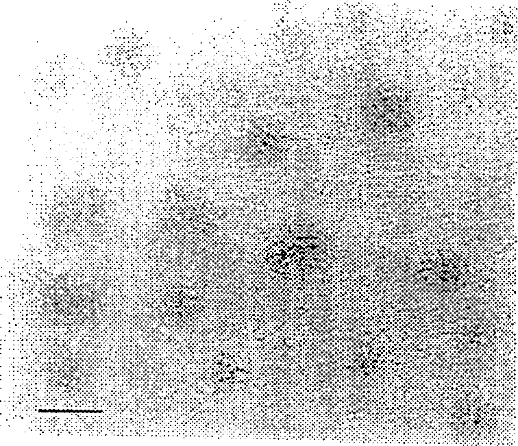
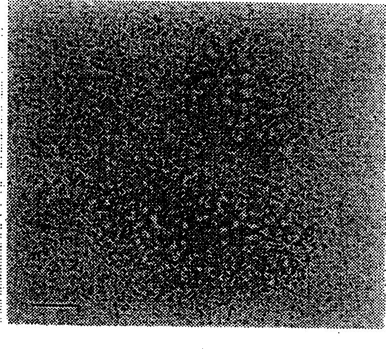
[0044] Figure 1a and 1b Chicken EG cell colonies after 3 passages are shown on chicken embryonic fibroblasts (CEF) ( Figure 1a : ruler, 50 μm; Figure 1b : scale bar, 25 μm). The morphology of chicken EG cells is slightly different from that of mouse ES living EG cells. Almost all chicken EG cell colonies were circular without firm borders with the CEG feeder layer. In contrast to mouse ES or EG cells, chicken EG cells do not pack tightly together; therefore individual member cells are not difficult to distinguish. The morphology of the colonies is multilayered with well-defined boundaries. Chicken EG cells have a large nucleus and a relatively small amount of cytoplasm, but their nucleoli are inconspicuous. Example 3: Characteristics of EG cells
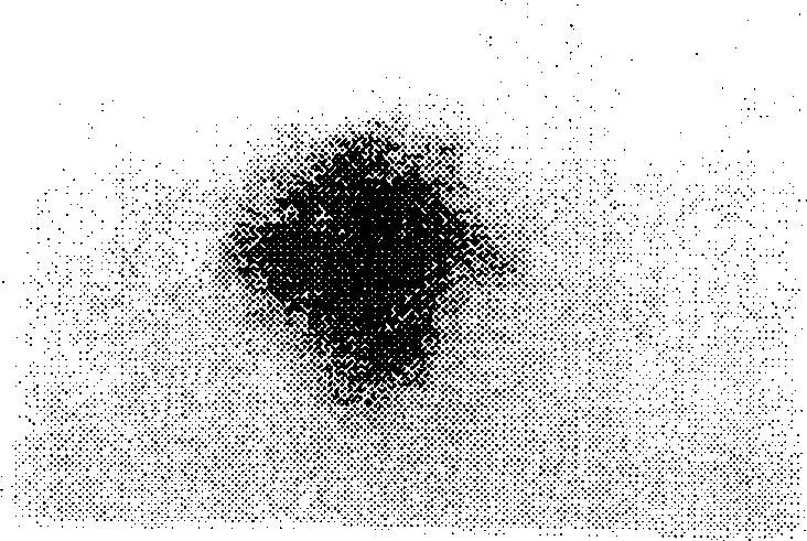
[0045] To determine whether the pluripotency of EG cells has characteristic properties of pluripotent cells, the presence of glycogen and SSEA-1 epitopes, their alkaline phosphatase activity and their ability to proliferate a...
PUM
 Abstract
Description
Claims
Application Information
 - R&D
- Intellectual Property
- Life Sciences
- Materials
- Tech Scout
- Unparalleled Data Quality
- Higher Quality Content
- 60% Fewer Hallucinations
Browse by: Latest US Patents, China's latest patents, Technical Efficacy Thesaurus, Application Domain, Technology Topic, Popular Technical Reports.
© 2025 PatSnap. All rights reserved.Legal|Privacy policy|Modern Slavery Act Transparency Statement|Sitemap|About US| Contact US: help@patsnap.com


