Allogenic tissue engineered cartilage and its application

A tissue engineering and cartilage technology, applied in the fields of medicine and biomedical engineering, can solve the problems of lack of cartilage formation ability, limited application, loss of ability to secrete cartilage matrix, etc.

Inactive Publication Date: 2004-02-25
上海第二医科大学附属第九人民医院
View PDF0 Cites 6 Cited by
  • Summary
  • Abstract
  • Description
  • Claims
  • Application Information

AI Technical Summary

Problems solved by technology

[0008] One of the important conditions for the construction of tissue-engineered cartilage tissue is to obtain a large number of functional and viable chondrocytes. Studies have shown that primary monolayer cultured chondrocytes can continue to express specific type II collagen for several days to several weeks, but In the subsequent subculture, chondrocytes undergo "dedifferentiation", that is, transform into fibroblast-like cells, begin to express type I and type III collagen, lose the ability to secrete cartilage matrix, and have no chondrogenic ability after implantation in the body, so It is difficult to obtain a large number of chondrocytes with normal functions after a small amount of autologous tissue is cultured and expanded in vitro, which limits its clinical application

Method used


Image

  • Allogenic tissue engineered cartilage and its application
  • Allogenic tissue engineered cartilage and its application
  • Allogenic tissue engineered cartilage and its application

Examples


preparation example Construction

[0047] The preparation method of the tissue engineered cartilage of the present invention is simple and convenient, and only needs to mix a certain amount of said chondrocytes with pharmaceutically acceptable biodegradable materials.

[0048] The tissue-engineered cartilage formed by the method of the present invention is divided into two types, one is a compound composed of chondrocytes and injectable biomaterials, and the compound can be directly injected into various parts of the body such as subcutaneous tissue and cartilage defects. The other is a compound of chondrocytes and non-injectable, which can be directly implanted into various parts of the body such as subcutaneous tissue and cartilage defects.

[0049] The advantages of the present invention are:

[0050] (1) Solved the problem of insufficient source of seed cells for cartilage tissue engineering, and aborted embryos can provide a large source of chondrocytes;

[0051] (2) Compared with adult chondrocytes, embr...

Embodiment 1

[0055] Isolation and culture of human embryonic chondrocytes

[0056] The human fetus of spontaneous abortion, the gestational age is about 5 months; within 10 hours after delivery. Separation of articular cartilage by aseptic operation Cut the harvested cartilage into pieces about 2mm×2mm in size, place them in a 15ml centrifuge tube, wash them twice with PBS, mix them with 15mg of type II collagenase per 1g of cartilage tissue, and use serum-free Adjust the concentration of type II collagenase to 1 mg / ml in F-12 medium. Digest in a constant temperature shaker at 37°C for 16 hours, filter with 150-mesh stainless steel mesh, centrifuge at 1500r / min, remove the supernatant, wash twice with PBS, resuspend chondrocytes in F-12 conditioned medium, stain with trypan blue, and count under a microscope , Cell viability > 85% can be used for tissue construction.

[0057] The yield of chondrocytes isolated from aborted fetal articular cartilage was 2×10 7 -4×10 7 One / gram human fet...

Embodiment 2

[0059] Proliferation of human embryonic chondrocytes in vitro

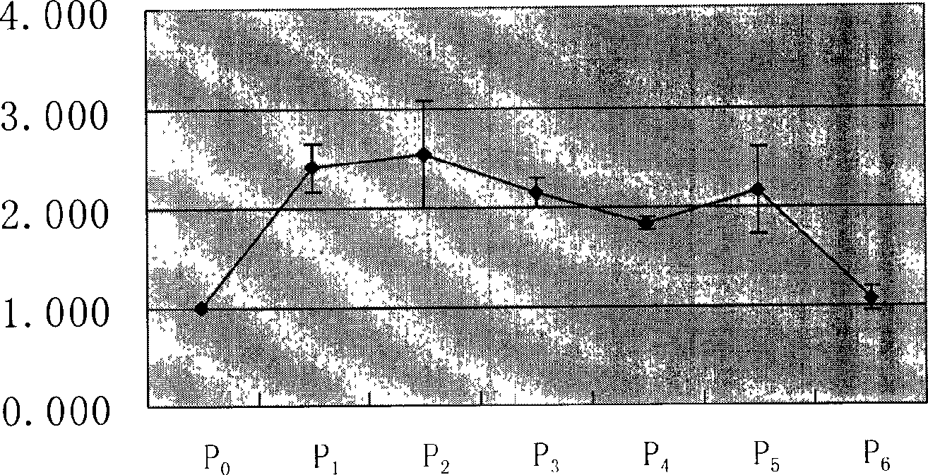
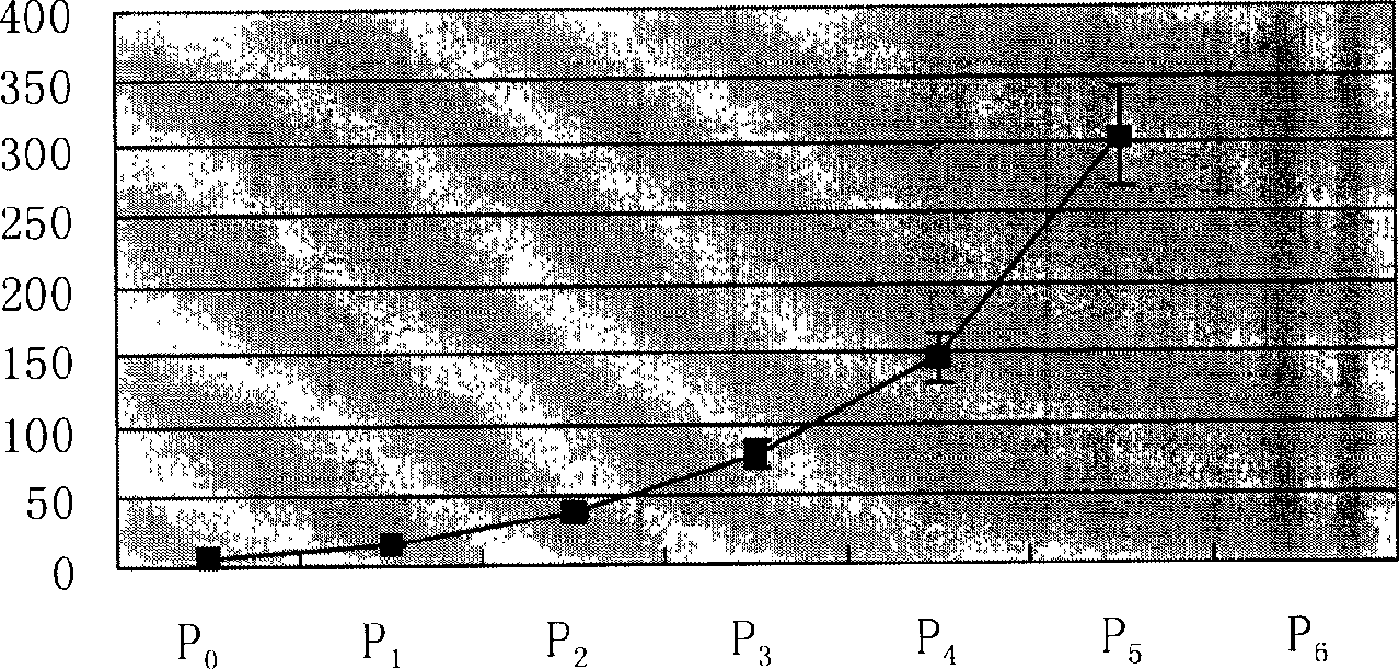
[0060] The number of cells obtained after the cells of each generation are expanded by culture, divided by the number of cells inoculated before culture, is the proliferation rate of the cells of this generation; the proliferation rate curve is drawn according to the proliferation rates of cells of different generations. Assuming that the number of primary cells is 1, according to the previously calculated proliferation rate, calculate the number of cells that can be obtained in each passage after expansion, and then draw the cell proliferation curve.

[0061] The result is as figure 1 and figure 2 As shown, when the human fetal chondrocytes were subcultured in vitro, the primary generation hardly proliferated, the first generation began to proliferate, and the proliferation rate was the highest at the second and third passages; the continuous passage to the sixth passage lost the ability to proliferate.


PUM


Abstract

The present invention provides an allogenic tissue engineered cartilage graft, it includes: (a) pharmaceutically acceptable bio-degradable material; and (b). cartilage cell, the described cartilage cell is one externally-cultured form primary culture to sixth generation. The invention can be used for effectively curing chondropathy. The invention also provides its preparation and application.

Description

technical field [0001] The present invention relates to the fields of medicine and biomedical engineering, and more particularly to tissue engineered cartilage and methods and uses thereof. Background technique [0002] Cartilage tissue, as a connective tissue, mainly plays the role of supporting, protecting and dispersing stress. Histologically, it can be divided into three types: hyaline cartilage (articular cartilage, costal cartilage, nasal cartilage, tracheal cartilage), elastic cartilage (epiglottic cartilage, ear cartilage), and fibrocartilage (meniscus). Histologically, they are single tissues containing only chondrocytes, without blood vessels inside, and their nutrition mainly depends on the penetration of tissue fluid. [0003] Structurally, chondrocytes are encased in cartilage lacunae, surrounded by a thick cartilage capsule that acts as a natural barrier to separate chondrocytes from surrounding tissue. Therefore, cartilage tissue has weak antigenicity, is no...

Claims


Application Information

Patent Timeline
IPC IPC(8): A61L27/00C12N5/073C12N5/077
Inventor 曹谊林刘伟崔磊
Owner 上海第二医科大学附属第九人民医院
Why Eureka
Try Eureka