Biological tissue refractivity space distribution function detecting method

A technology of spatial distribution function and biological tissue, which is applied in the field of detection of optical parameters of biological tissue, and can solve the problems of the difficulty of spatial distribution function of optical transmission parameters and the limitation of measurement accuracy.

Inactive Publication Date: 2005-02-16
BEIJING JIAOTONG UNIV
View PDF0 Cites 8 Cited by
  • Summary
  • Abstract
  • Description
  • Claims
  • Application Information

AI Technical Summary

Problems solved by technology

[0010] In order to overcome the difficulties of the existing measurement methods on the spatial distribution function of the optical transmission parameters of biological tissue, and the limitations of the turbidity and high scattering characteristics of biological tissue on the measurement accuracy of visible and near-infrared light experiments, the present invention treats biological tissue from the perspective of photonic crystals, A detection method for the spatial distribution function of the refractive index of biological tissue based on photonic crystal theory is proposed
[0011] The technical scheme adopted by the present invention to solve the problems of the existing biological tissue optical transmission parameter spatial distribution function measurement method is as follows

Method used


Image

  • Biological tissue refractivity space distribution function detecting method
  • Biological tissue refractivity space distribution function detecting method
  • Biological tissue refractivity space distribution function detecting method

Examples


Embodiment 1

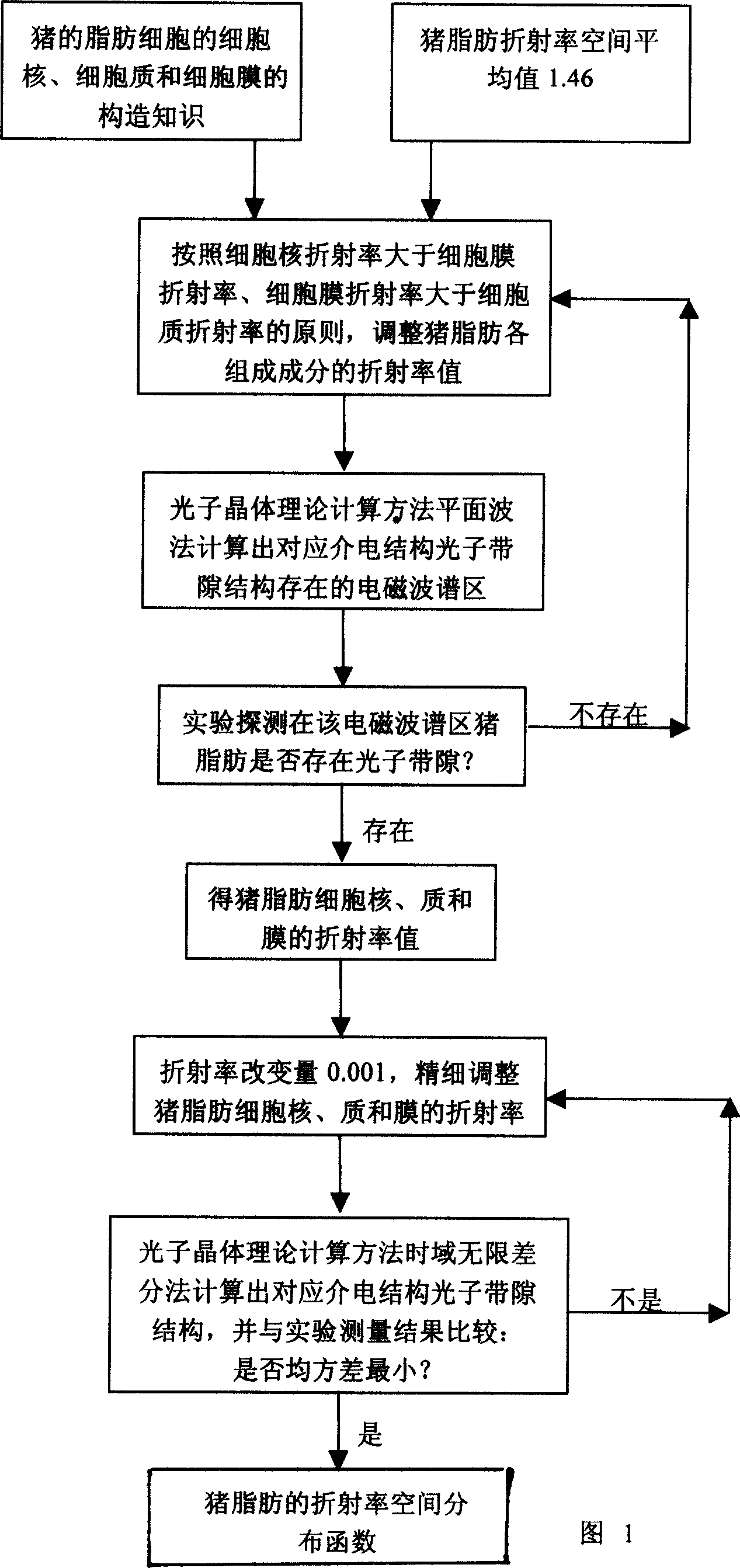
[0025] Embodiment 1: Embodiment 1 of the method for detecting the spatial distribution function of the refractive index of biological tissue, the flow chart is shown in FIG. 1 .

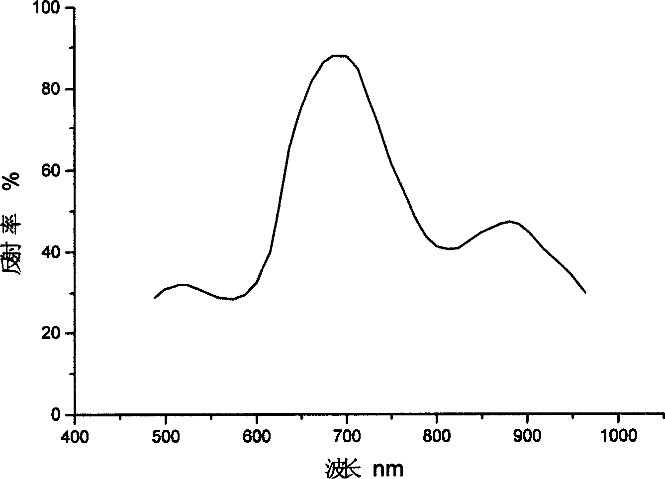
[0026] Step 1: Determine a biological tissue object as pig fat, according to the tissue structure of the nucleus, cytoplasm and cell membrane of pig fat and the spatial average value of the refractive index of 1.46, according to the structure that the refractive index of the nucleus is greater than the refractive index of the cell membrane, and the refractive index of the cell membrane is greater than the refractive index of the cytoplasm, Adjust the spatial distribution of the refractive index in pig fat, and use the photonic crystal theoretical calculation method to estimate the electromagnetic spectrum region where the photonic bandgap structure of pig fat is located; figure 2 Shown is the result of one of these calculations.

[0027] Step 2: In the electromagnetic spectrum region, use experimenta...

Embodiment 2

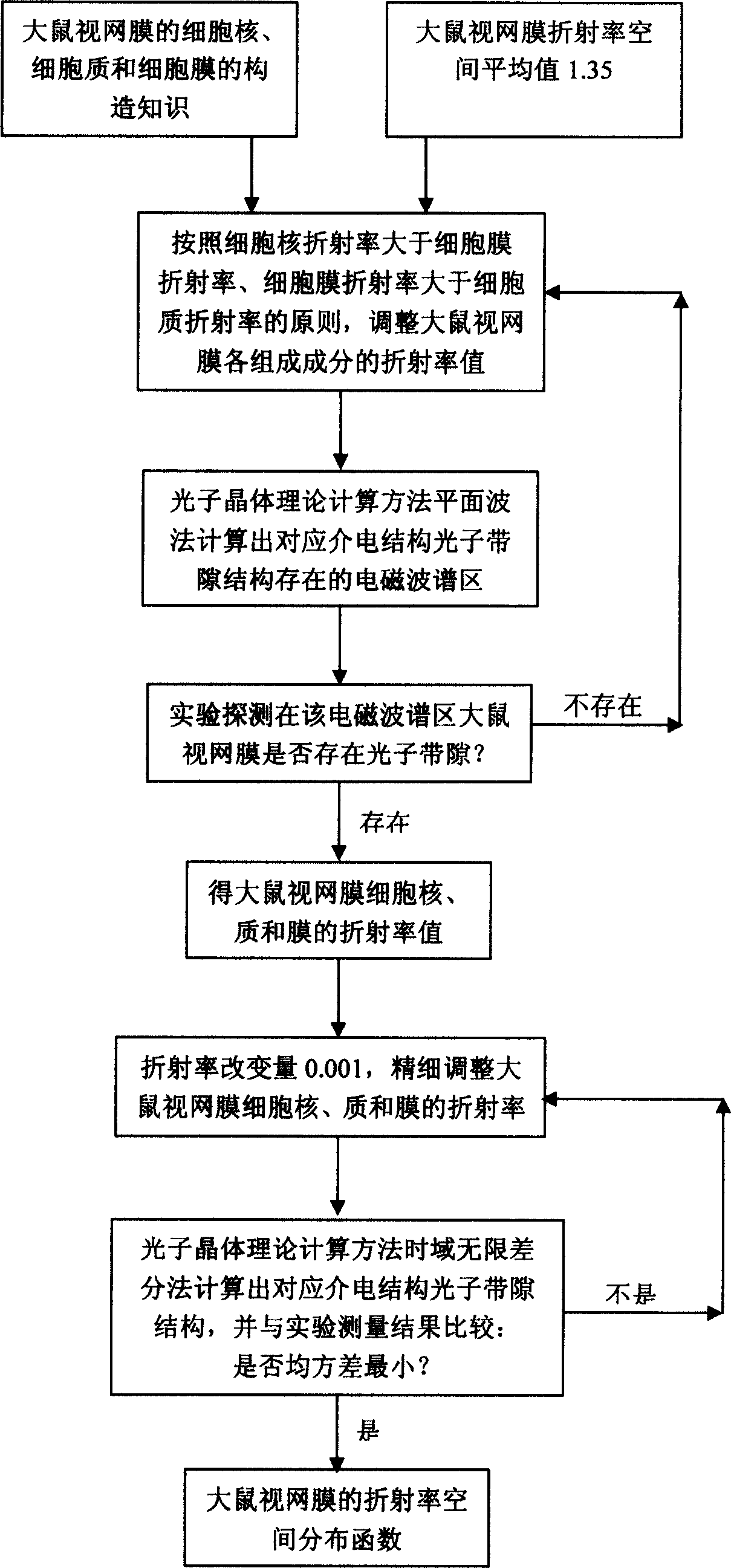
[0034] The second embodiment of the method for detecting the spatial distribution function of the refractive index of biological tissue is shown in FIG. 5 .

[0035] Step 1: Determine that a biological tissue object is the retina of a rat. According to the tissue structure of the nucleus, cytoplasm and cell membrane of the rat retina and the spatial average value of the refractive index of 1.35, the refractive index of the nucleus is greater than the refractive index of the cell membrane, and the refractive index of the cell membrane is greater than the refractive index of the cytoplasm Based on the framework, the spatial distribution of the refractive index in the rat retina is adjusted, and several electromagnetic spectrum regions where the photonic bandgap structure of the rat retina is located are estimated by using the photonic crystal theoretical calculation method;

[0036] Step 2: In the electromagnetic spectrum region, use experimental methods including absorption spec...

Embodiment 3

[0043] With the steps of Example 1 or Example 2, the difference is that biological tissues include fat, muscle and connective tissue of mammals or non-mammals, such as fat, muscle and connective tissue of mammalian people or cattle or cats or dogs, such as Fat, muscle and connective tissue of non-mammalian eagles or fish or chickens and crocodiles; knowledge of biological tissue structure is the knowledge of the composition of biological tissues, as well as the knowledge of the geometric dimensions of the nucleus, cytoplasm and cell membrane, and the distance between cells.


PUM


Abstract

This is a space-distributed function testing method for the biological organism refractive index. This method use the photon theory calculated method to calculate the biological organism ribbon chink structure, by the treatment of photon crystal visual angle to treat the biological organism. To adjust the refractive index of biological organism to approach the experiment surveying result of the biological organism ribbon chink structure, then to avoid away the muddy and high scattering characteristic of the biological organism to influence the survey precision, to raise the space distributed index of biological organism refractive index.

Description

technical field [0001] The invention relates to a detection method for optical parameters of biological tissue, in particular to a detection method for the spatial distribution function of the refractive index of biological tissue. Background technique [0002] The problem of how light propagates in biological tissues is a bottleneck problem that hinders the development of hot biomedical technologies such as optical coherence tomography, tumor photodynamic therapy and laser-induced thermotherapy. The advancement of these techniques depends on the accurate measurement of the spatial distribution functions of the optical parameters of biological tissues. [0003] The importance and complexity of light transport properties in biological tissues has led to a new branch of discipline - tissue optics formation. In 1988, the OSA annual meeting in the United States set up a special topic to discuss the optical properties of mammalian tissues, marking the rise of tissue optics. At ...

Claims


Application Information

Patent Timeline
Patent Type & Authority Applications(China)
IPC IPC(8): G01N21/25G01N23/02
Inventor 吴重庆盛新志
Owner BEIJING JIAOTONG UNIV
Why Eureka
Try Eureka