Tumor tomography method and device with phonochemistry luminance provoking function by focused ultrasound

A technology of sonochemiluminescence and focused ultrasound, applied in the field of medical tomography and medical devices, can solve the problems of inability to overcome fluorescence interference, skin phototoxic side effects, and high diagnostic costs, and achieves reduction of photochemical toxic side effects and high penetrating ability. Sensitivity, diagnostic signal-to-noise ratio improvement effect

Inactive Publication Date: 2005-09-07
SOUTH CHINA NORMAL UNIVERSITY
View PDF0 Cites 4 Cited by
  • Summary
  • Abstract
  • Description
  • Claims
  • Application Information

AI Technical Summary

Problems solved by technology

In view of the fact that the etiology and pathogenesis of tumors are not clear at present, the treatment methods related to tumors cannot fundamentally solve the problem.
Therefore, the diagnosis of malignant tumors in medicine must rely on the comprehensive application of clinical diagnosis, instrumental diagnosis (such as CT, MRI, B-ultrasound, etc.), laboratory (tumor markers, genetic testing, etc.) The means to make correct judgments, the cost of diagnosis is extremely high, and early tumor diagnosis cannot be achieved
[0004] Biomedical photonics imaging technology has the advantages of no damage to the human body, high sensitivity, and functional imaging, but the disadvantages are small penetration depth and strong biological autofluorescence interference
On the other hand, the penetration performance of ultrasound in the body is very good, but the current imaging technology based on ultrasound (such as B-ultrasound) can only do interface reflection imaging, and the sensitivity is low
For the optical imaging of tumor tissue, the laser-induced drug fluorescence imaging method is generally used. Because it uses a short-wavelength excitation light source, it has problems such as tissue autofluorescence interference and small penetration depth that cannot be overcome. In addition, when using this method, the Skin phototoxic side effects will inevitably occur, which will cause greater damage to patients

Method used


Image

  • Tumor tomography method and device with phonochemistry luminance provoking function by focused ultrasound
  • Tumor tomography method and device with phonochemistry luminance provoking function by focused ultrasound
  • Tumor tomography method and device with phonochemistry luminance provoking function by focused ultrasound

Examples


Embodiment Construction

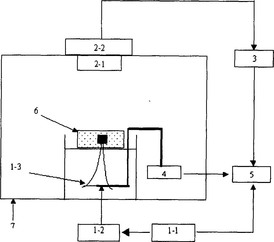
[0038] figure 1 It is a structural block diagram of the tumor acoustochemiluminescence tomography device of the present invention, consisting of figure 1It can be seen that the device is composed of an ultrasonic generating component 1, a light receiving component 2, an analog-to-digital converter 3, a three-dimensional platform 4, and a computer 5, wherein the ultrasonic generating component 1 is composed of a function generator 1-1, a power amplifier 1-2, a focused ultrasonic The transducers 1-3 are electrically connected in sequence; the light receiving component 2 is formed by connecting the camera lens 2-1 and the detector 2-2; the ultrasonic generating component 1, the three-dimensional platform 4 are electrically connected with the computer 5, and the light receiving component 2 , the analog-to-digital converter 3 is electrically connected to the computer 5 in turn; 6 is the measured organism (or tissue), and 7 is the darkroom. Each component is selected to connect to ...


PUM


Abstract

The invention relates to a sonochemistrical luminous tomography method for tumour, which is that after distributing of sonosensitizer in tumour group, generating active oxygen-derived free radicals by scanning point by point and sensitizing used focusing ultrasound; translating the active oxygen-derived free radicals generated during sonosensitizing process real time and directly by chemical luminous agents into photons and radiating out; based on that, simulating the imaging of tumour group by optics detection technique. The apparatus comprises ultrasound producing groupware, light receiving groupware, A/D converter, triaxiality scanning device and computer; wherein ultrasound producing groupware and triaxiality scanning device link to computer, light receiving groupware and A/D converter link to computer in turn. The invention combines strong penetrating ability of focusing ultrasound to body and the advantages of high sensitivity and intactness of optics imaging, brings no radioactive damage to organism.

Description

technical field [0001] The present invention relates to medical tomography and medical devices. In more detail, a focused ultrasound-induced acoustochemiluminescence method for tumor tomography. [0002] The invention also relates to a device for carrying out the above-mentioned method. Background technique [0003] Tumor is a common and frequently-occurring disease, among which malignant tumor is a kind of disease that seriously endangers human health, and its mortality rate ranks second only to cardiovascular disease. In view of the fact that the etiology and pathogenesis of tumors are not clear at present, the treatment methods related to tumors cannot fundamentally solve the problem. Therefore, the diagnosis of malignant tumors in medicine must rely on the comprehensive application of clinical diagnosis, instrumental diagnosis (such as CT, MRI, B-ultrasound, etc.), laboratory (tumor markers, genetic testing, etc.) The means to make a correct judgment, the diagnosis co...

Claims


Application Information

Patent Timeline
Patent Type & Authority Applications(China)
IPC IPC(8): A61B8/08A61B8/13
Inventor 邢达谭毅
Owner SOUTH CHINA NORMAL UNIVERSITY
Why Eureka
Try Eureka