Nanofiber-based graft for heart valve replacement and methods of using the same

a technology of nanofibers and heart valves, applied in the field of electro-optical nanofiber biomaterials, can solve the problems of life-threatening dysfunctional heart valves, early valve degeneration, and need life-long anti-coagulant therapy, and achieve the effects of reducing thrombogenic potential, reducing thrombogenic potential, and improving thrombolytic potential

Inactive Publication Date: 2019-03-05
WAKE FOREST UNIV HEALTH SCI INC
View PDF60 Cites 21 Cited by
  • Summary
  • Abstract
  • Description
  • Claims
  • Application Information

AI Technical Summary

Benefits of technology

[0008]In another aspect, the present invention encompasses a porous electrospun graft material which is readily configured to function as a tissue replacement. The graft comprises a collagen, a fibroin, and a hemocompatible synthetic elastomer. In one embodiment, the tissue to be replaced is heart tissue. In another embodiment, the graft is molded in the shape of a heart valve.
[0011]The present invention provides electrospun grafts having superior mechanical properties, low degradation, and reduced thrombogenic potential compared to primarily collagen-based materials.

Problems solved by technology

Dysfunctional heart valves are life-threatening as the diseased valvular tissues are unable to perform the normal physiological requirements.
Commonly used mechanical heart valves have adequate durability but are often thrombogenic and require life-long anti-coagulant therapy.
Bioprosthetic collagen-based tissue valves from porcine valves or bovine pericardium mimic the anatomy of native valves, however, early valve degeneration, and 50% postoperative failure occur within 12-15 years.

Method used


Image

  • Nanofiber-based graft for heart valve replacement and methods of using the same
  • Nanofiber-based graft for heart valve replacement and methods of using the same
  • Nanofiber-based graft for heart valve replacement and methods of using the same

Examples


example 1

Composite Characterization

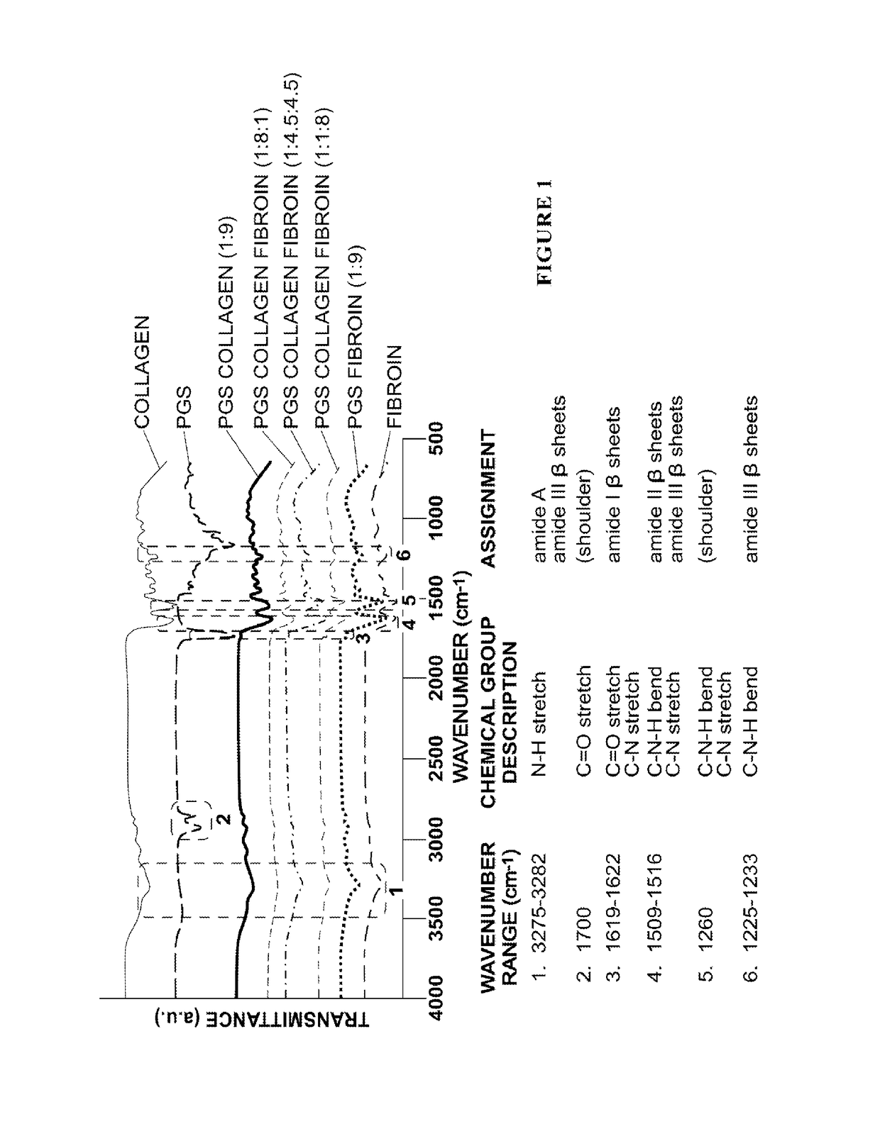
[0092]The blending of materials in composite nanofibers is initially demonstrated. For the extracted fibroin, FTIR spectroscopy indicated amide I, II, III groups were in the β sheet conformation based on wavenumbers corresponding to the carbonyl stretchs 1619-1622 cm−1, 1509-1516 cm−1, and 1225-1233 cm−1 respectively [23-24]. The wavenumber ranging from 3275-3282 cm−1 was indicative of the —N—H stretching vibration shown as a broad peak for amide A. An absorption peak of 1700 cm−1 was assigned to be the C═O stretch in amide I β sheets, and 1225-1233 cm−1 referred to the C—N stretch and C—N—H bend in amide III β sheets structure (Hu et al. 2006; Hayashi et al. 2007). Two strong peaks were shown at the regions of 1619-1622 cm−1 for C═O stretch of amide I and 1509-1516 cm−1 for the C—N stretch and C—N—H bend for amide II (Horan et al. 2005). (FIG. 1). Type I collagen had a characteristic broad peak at the absorption of 3275 cm−1 which was indicative for the —O...

example 2

Fiber Morphology

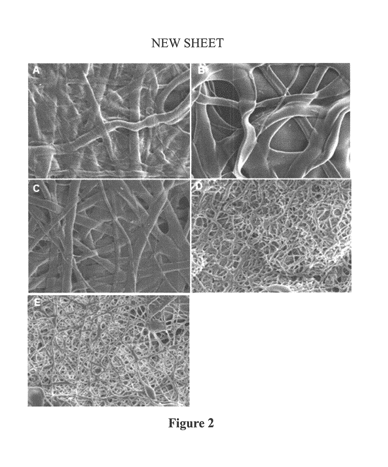
[0093]Fiber diameters ranged from 694 to 4577 nm (Table 1). In general, thinner and more rounded fibers were observed for the electrospun mats with higher fibroin content (90% and 80%) as compared to the thicker and more flat fiber of electrospun mats with high proportions of collagen (90%, 80%, and 45%). The interconnected fiber network structures of electrospun mats at various collagen, fibroin and PGS weight ratios were compared after crosslinking using scanning electron microscopy (SEM) (FIG. 2).

[0094]

TABLE 1Fiber Diameters in Mats of Different CompositionsSample TypeFiber Diameters (nm)Collagen:PGS (9:1)2067 ± 168Collagen:Fibroin:PGS (8:1:1)4577 ± 697Collagen:Fibroin:PGS (4.5:4.5:1)2952 ± 240Collagen:Fibroin:PGS (1:8:1)784 ± 77Fibroin:PGS (9:1)694 ± 43All values represent means ± SEM;The fiber diameters were measured from 16 randomly selected fibers of two representative SEM pictures. The measurements are presented as mean ± standard error of the mean.

example 3

Thermal Transition Analysis of Electrospun Mats

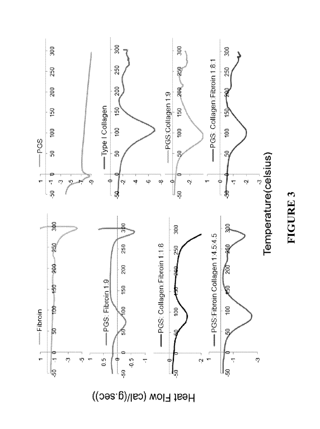
[0095]Results of DSC scan were analyzed to determine thermal transition temperatures of electrospun mats (FIG. 3). By incorporating an increasing amount of fibroin, a shift of thermal transition temperature to higher range was observed. The electrospun composite materials exhibit much higher thermal transition temperatures as compared to PGS alone. Results suggest the electrospun mats made from collagen, silk fibroin, and PGS composites were thermally stable for in vivo application.


PUM

PropertyMeasurementUnit
diameteraaaaaaaaaa
diameteraaaaaaaaaa
thickaaaaaaaaaa

Abstract

Nanofiber-based biomaterials containing fibroin for wound repair and tissue replacement and, more particularly, heart valve replacement.

Description

REFERENCE TO RELATED APPLICATIONS[0001]This application is a 371 application of International Application No. PCT / US2013 / 066747, filed Oct. 25, 2013, which claims the benefit of U.S. Provisional Application No. 61 / 718,991, filed Oct. 26, 2012, the entirety of which are incorporated by reference herein.FIELD OF THE INVENTION[0002]The present invention relates generally to electrospun nanofiber-based biomaterials useful for wound repair and tissue replacement and, more particularly, but not exclusively to electrospun composite materials comprising fibroin for tissue engineering applications, particularly heart valve replacement.BACKGROUND OF THE INVENTION[0003]Severe valvular heart disease (VHD) affects 1 out of 40 adults in the United States, and is responsible for approximately 28,000 deaths per year. Aortic valves consist of 3 film-like cusps with an average thickness of 300-700 μm. The main composition of the valvular extracellular material (ECM) is type I collagen and elastin in ...

Claims


Application Information

Patent Timeline
Patent Type & Authority Patents(United States)
IPC IPC(8): A61F2/24C08J9/00A61L27/56A61L27/54A61L27/26A61F2/82A61K9/70C08L89/00C08L89/06
CPCA61F2/2412A61F2/2415A61F2/82A61K9/70A61L27/26A61L27/54A61L27/56C08J9/0061C08L89/00C08L89/06C08L31/06Y10T428/298C08L2205/03C08L2205/02A61L2300/414A61L2400/12A61L2430/20C08J2389/00C08J2389/06C08J2467/02C08J2489/00C08J2489/06C08L2201/06C08L2203/02C08L2203/12
Inventor WAGNER, WILLIAM D.LEVI, NICOLEWANG, RUIARGENTA, LOUIS C.MORYKWAS, MICHAEL J.
Owner WAKE FOREST UNIV HEALTH SCI INC
Why Eureka
Try Eureka