
Antigen detection method which uses lectin and comprises enzyme treatment step

a detection method and enzyme treatment technology, applied in the field of antigen detection methods using lectins, can solve the problems of difficult preparation of antigens against sugar chains, low specificity of lectins, and low binding activity of lectins, and achieve the effects of improving detection sensitivity and quantitative performance, simple technique, and simple constitution

Active Publication Date: 2020-01-07
OTSUKA PHARM CO LTD
View PDF90 Cites 0 Cited by
  • Summary
  • Abstract
  • Description
  • Claims
  • Application Information

AI Technical Summary

Benefits of technology

[0026]According to the present invention, an antigen detection method which can suppress an increase in the background of a measurement system and improve the detection sensitivity and quantitative performance for a sugar chain on an antigen to be detected with a lectin by a simple technique (simple constitution) in an antigen detection system using a lectin, as well as a detection kit can be provided.
[0027]Further, since the above-described effects can be attained only by incorporating an enzyme into a reagent in a sandwich assay system, the antigen detection method of the present invention is a simple technique (simple constitution) and can be effectively applied to diagnosis scene.

Problems solved by technology

This is because it is very difficult to prepare an antigen against a sugar chain and such an antigen is hardly available.
However, lectins have such drawbacks of having lower binding activity and specificity than antibodies.
For example, N-acetylgalactosamine is known as a main binding sugar chain in Wisteria floribunda lectin (WFA); however, since N-acetylgalactosamine shows slight binding with galactose, it may non-specifically react with a galactose residue if present in a reaction system.
However, although this technique is effective when the subject protein is purified to a certain extent, since it shows markedly reduced performance in terms of sensitivity and quantitative capacity in a system that contains a large amount of contaminants, such as glycoproteins and glycolipids that are not measurement subjects and included in blood, urine and the like to be used as a sample in ordinary disease diagnosis, it is very difficult to perform an analysis using this technique.
However, it is an extremely rare case where these background-suppressing measures show a drastic effect in quantitative analysis using a lectin for detection, and there is also a problem that the search and examination of subject-suitable blocking agent require tremendous man-hours.

Method used


Image

  • Antigen detection method which uses lectin and comprises enzyme treatment step
  • Antigen detection method which uses lectin and comprises enzyme treatment step
  • Antigen detection method which uses lectin and comprises enzyme treatment step

Examples


preparation example 1

[Anti-PSA Antibody-Solid Phased SAM Film (Substrate)]

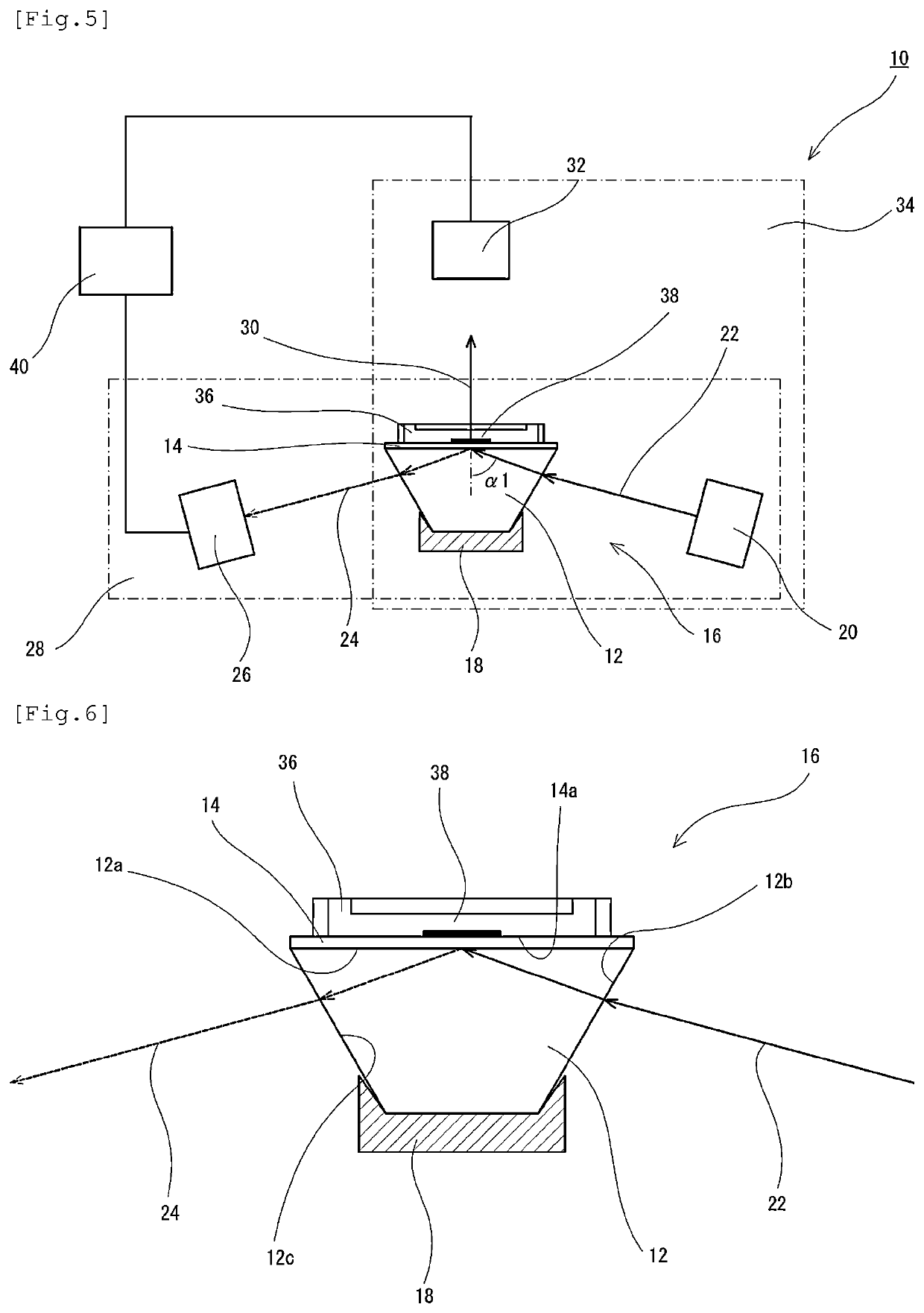
[0146]In the external flow path to which the plasmon excitation sensor was connected as described above, ultrapure water and then phosphate buffered saline (PBS) were circulated for 10 minutes and 20 minutes, respectively, using a peristaltic pump at a room temperature (25° C.) and a flow rate of 500 uL / min, thereby equilibrating the surface of the plasmon excitation sensor.

[0147]Subsequently, after transferring and circulating 5 mL of a phosphate buffered saline (PBS) containing 50 mM of N-hydroxysuccinic acid imide (NHS) and 100 mM of water-soluble carbodiimide (WSC) for 20 minutes, 2.5 mL of an anti-PSA monoclonal antibody solution (No. 79, 2.5 mg / mL; manufactured by Mikuri Immunolaboratory, Ltd.) was circulated for 30 minutes to solid-phase the antibody on the SAM film, thereby preparing an anti-PSA antibody-solid phased SAM film.

[0148]It is noted here that a non-specific adsorption-inhibiting treatment was performed in the fl...

preparation example 2

[Anti-AFP Antibody-Solid Phased SAM Film (Substrate)]

[0150]An anti-AFP antibody-solid phased SAM film was prepared in the same manner as in Preparation Example 1, except that an anti-AFP monoclonal antibody (1D5, 2.5 mg / mL; manufactured by Japan Clinical Laboratories, Inc.) was used in place of the anti-PSA antibody.

Preparation of Labeled Lectins

production example 1

[Fluorescently-Labeled WFA Lectin]

[0151]A fluorescently-labeled WFA lectin was produced using a fluorescent substance labeling kit, “Alexa Fluor (registered trademark) 647 Protein Labeling Kit” (manufactured by Invitrogen Corp.). Then, 100 μg equivalent of a WFA lectin (L-1350, manufactured by Vector Laboratories, Inc.), 0.1M sodium bicarbonate and Alexa Fluor 647 reactive dye were mixed and allowed to react at room temperature for 1 hour. Subsequently, the resultant was subjected to gel filtration chromatography and ultrafiltration, thereby removing Alexa Fluor 647 reactive dye that was not utilized in labeling to obtain a fluorescently-labeled WFA lectin. Thereafter, the absorbance was measured to determine the concentration of the labeled lectin.


PUM

PropertyMeasurementUnit
chemiluminescent substanceaaaaaaaaaa
stabilityaaaaaaaaaa
concentrationaaaaaaaaaa

Abstract

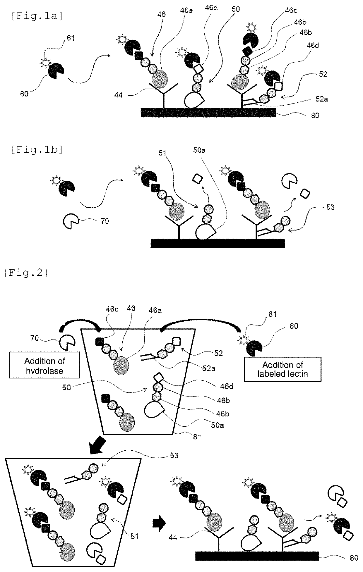
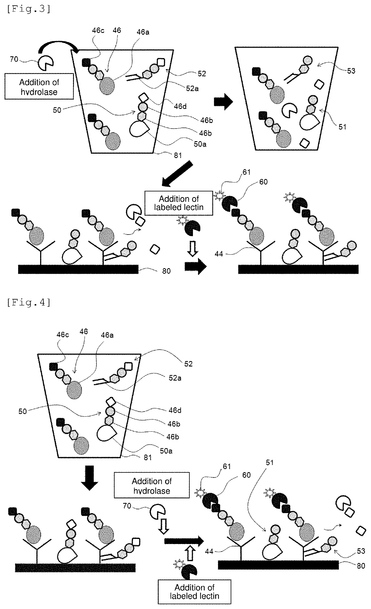
An antigen detection method detects an antigen having a specific sugar chain in a sample with a lectin that binds to plural kinds of sugar chains including the specific sugar chain. The detection method includes: a first step of bringing the lectin into contact with the sample; a second step of bringing a glycohydrolase capable of cleaving at least one kind of sugar chain to which the lectin can bind into contact with the sample, the at least one kind of sugar chain excluding the specific sugar chain among the plural kinds of sugar chains; and a step of detecting the antigen bound with the lectin after the first and second steps.

Description

TECHNICAL FIELD[0001]The present invention relates to an antigen detection method using a lectin. More particularly, the present invention relates to a high-sensitivity and low-noise antigen detection method using a lectin, the method comprising the step of subjecting a sample to an enzyme treatment.BACKGROUND ART[0002]In order to allow proteins assuming a principal role in the biological life functions to orderly exert their functions in the cellular world, post-translation modifications including glycosylation play an extremely important role. In recent years, it has been gradually revealed that most in vivo proteins undergo modifications with sugar chains and that those sugar chains attached to proteins play important roles in various aspects of biological phenomena such as viral infection, protozoan parasitism and infection, binding of toxins, binding of hormones, fertilization, development and differentiation, protein stability, cancer cell metastasis, and apotosis.[0003]It is ...

Claims


Application Information

Patent Timeline
Patent Type & Authority Patents(United States)
IPC IPC(8): C12Q1/34G01N33/566G01N33/53
CPCG01N33/566C12Q1/34G01N33/5308G01N2333/42G01N2333/4724
Inventor KANEKO, TOMONORIIDE, YOUICHI
Owner OTSUKA PHARM CO LTD
Learn More