Integrated intraoperative diagnosis and thermal therapy system

a technology of thermal therapy and intraoperative diagnosis, applied in the field of multimodal medical devices and methods, can solve the problems of increasing morbidity, increasing the cost, and inability to provide timely treatment, and achieve the effects of reducing cost, improving accuracy and success rate of procedures, and accurate diagnosis and treatmen

Active Publication Date: 2020-02-25
SHANGHAI JIAOTONG UNIV +1
View PDF14 Cites 2 Cited by
  • Summary
  • Abstract
  • Description
  • Claims
  • Application Information

AI Technical Summary

Benefits of technology

We have invented a multimodal medical device for the diagnosis and treatment of cancer, cardiac and cardiovascular disease. This system combines thermal therapy with several tomographic imaging modes, such as optical coherence tomography (OCT), ultrasound imaging (US), photoacoustic (PA) imaging, fluorescence imaging and thermal imaging guidance to provide accurate diagnosis and treatment of cancer and cardiac and cardiovascular diseases. The current illustrated embodiments of the invention, which can diagnose and treat the disease simultaneously, will greatly decrease cost and provide more timely treatment. Our illustrated embodiments can be applied for the diagnosis and treatment of several kinds of deadly diseases, which include cardiac and cardiovascular diseases and cancer.
The illustrated embodiments of the invention further improve the treatment by incorporating dual or multiple balloons with the catheter. One balloon will be used for cryoplasty and / or thermal therapy and another balloon will be used for isolation. Under the guidance of the OCT image, the location of the plaque will be accurately diagnosed and located. Typical plaque only exits on one side of the vessel. With this arrangement, only the side with plaque will accept the cryoplasty therapy and the other side will not be affected by the cryoplasty therapy. This will increase the treatment accuracy and minimize unnecessary treatment of healthy tissue.

Problems solved by technology

Separate diagnosis and treatment increases the cost and cannot provide timely treatment, which may increase morbidity in some cases.
However, without an imaging guidance and a monitoring technique, the accuracy and success rate of a procedure is limited.
However, surgical bypass procedures are associated with periprocedural morbid events, such as wound infection.
Conventional PTA is limited by high rates of vascular recoil, dissection, and restenosis, especially in the cases of infrainguinal intervention.
Although stenting improved periprocedural success by successfully treating vascular recoil and dissection, it still produces unacceptably high restenosis rates and low long-term potency rates, especially for below-the-knee lesions.
Traditional angiography only shows an image of the lumen of the vessels; it is impossible for the angiographic image to tell exactly where the atherosclerotic plaque is located.
Recent work in IVUS backscatter analysis demonstrated the feasibility and limitation of IVUS to characterize specific lesions and identify plaques that lead to various clinical syndromes.
However, current IVUS has limited resolution and sensitivity to assess the thickness of thin fibrous caps and for plaque classifications.
However, this high cure rate is not possible in other cancer modalities yet.
One reason is the limited high resolution imaging methods for guiding treatment and monitoring treatment at internal body sites to ensure sufficient killing by the thermal treatment, the heating or freezing.
However, ultrasound / CT / MRI is limited by its resolution from which it is very difficult to define the fine boundary of the tumor and provide a clear monitoring of tissue deformation under temperature change.
Radiotherapy and chemotherapy are the standard treatments for lung cancer but have limited effectiveness in reopening obstructed airways.
Patients with obstructed airways are not considered suitable for surgery, the other standard treatment for lung cancer, either.
It is very challenging to kill tumor tissue without heavy bleeding or cuting important nerves by conventional surgery.

Method used


Image

  • Integrated intraoperative diagnosis and thermal therapy system
  • Integrated intraoperative diagnosis and thermal therapy system
  • Integrated intraoperative diagnosis and thermal therapy system

Examples


Embodiment Construction

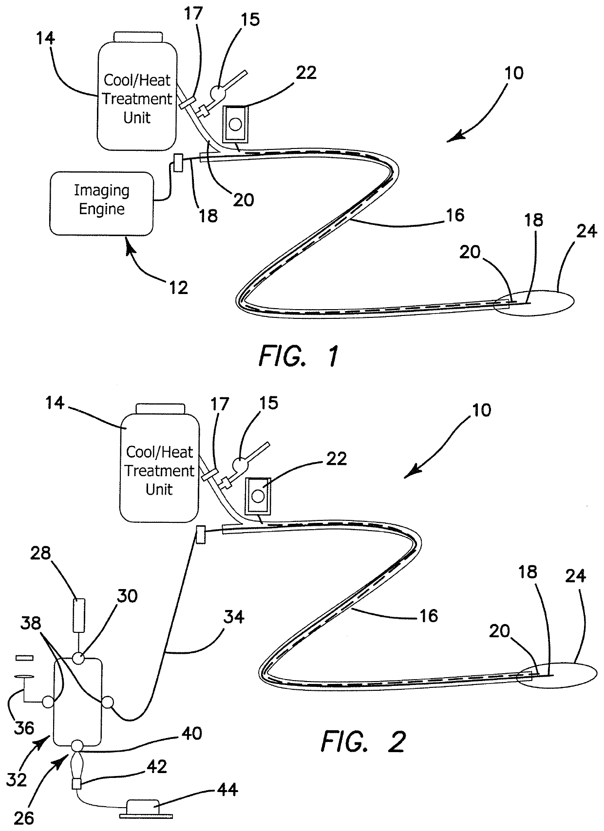
The disclosed multimodal system for the diagnosis and treatment of cancer and cardiac disease combines thermal therapy with tomographic imaging and thermal imaging guidance to provide accurate diagnosis and treatment of cancer and cardiac disease. With the help of several low cost imaging modalities, such as optical coherence tomography (OCT), ultrasound imaging, photoacoustic (PA) imaging, fluorescence imaging and thermal imaging, thermal therapy can be performed with much higher accuracy. The illustrated embodiment is employed in the following fields.

As shown in FIG. 1, the multimodal diagnosis and therapy system, generally denoted by reference numeral 10, includes an imaging system 12 and a cooling and / or heating unit or thermoplasty system 14. The imaging system 12 may include several imaging modualities such as OCT, ultrasound, photoacoustic, fluorescence and thermal imaging. These imaging modualities can also be integrated into a single catheter 16. The imaging catheter 16 and...


PUM


Abstract

A multimodal medical device for the diagnosis and / or thermal treatment of a disease includes an instrument for performing thermal therapy; and an imaging system having multiple tomographic imaging modes, including optical coherence tomography (OCT), ultrasound imaging, photoacoustic (PA) imaging, fluorescence imaging and / or thermal imaging to provide guidance for accurate diagnosis and treatment of the disease.

Description

RELATED APPLICATIONSThis application is related to provisional patent application, entitled, AN INTEGRATED INTRAOPERATIVE DIAGNOSIS AND THERAPY CATHETER SYSTEM Ser. No. 62 / 104,594, filed on Jan. 16, 2015, under 35 USC 119, which is incorporated herein by reference.BACKGROUND1. Field of the TechnologyThe invention relates to the field of multimodal medical devices and methods for the diagnosis and treatment of cancer, cardiac and cardiovascular disease.2. Description of the Prior ArtIn traditional cardiology and oncology, disease diagnosis and treatment procedures are usually separate. Separate diagnosis and treatment increases the cost and cannot provide timely treatment, which may increase morbidity in some cases. A system which can diagnose and treat diseases simultaneously will greatly decrease the cost and provide timely treatment, which may prevent death from the disease.Thermal therapy with its relative low cost, few side-effects and short procedure time has been widely used i...

Claims


Application Information

Patent Timeline
Patent Type & Authority Patents(United States)
IPC IPC(8): A61B18/14A61B18/18A61B18/02A61B18/00A61B8/08A61B18/24A61B5/00
CPCA61B18/1492A61B18/245A61B5/0066A61B5/0095A61B2018/00345A61B2018/1807A61B2018/0212A61B2018/00577A61B5/0071A61B8/08A61B2018/00982A61B18/1815A61B2018/1861A61F7/12A61F2007/126A61B5/0036A61B2018/00386A61N1/403A61B8/12A61B8/4416A61B8/445A61B5/6853A61B5/0084A61B5/0035A61N7/022A61B2018/00023A61B2018/0022A61B2018/00791A61B2090/3735A61B2090/378A61B2090/3782
Inventor CHEN, ZHONGPINGLIU, GANGJUNLI, JIAWENXU, LISA X.ZHANG, AILI
Owner SHANGHAI JIAOTONG UNIV
Why Eureka
Try Eureka