Vaccine formulation for ocular immunization

a technology for ocular immunization and vaccine formulation, applied in the field of ocular therapeutics, can solve the problems of affecting the quality of life of millions, ocular surface diseases, especially ocular surface infections, are globally under-recognized and neglected, and achieve the effect of increasing the uptake of corpuscular adjuvants

Active Publication Date: 2020-07-14
MEDIZINISCHE UNIVET WIEN
View PDF7 Cites 0 Cited by
  • Summary
  • Abstract
  • Description
  • Claims
  • Application Information

AI Technical Summary

Benefits of technology

[0020]Efficient delivery of protein antigens via the conjunctival route of immunization is of remarkable interest in order to both (i) mimic the bacterial infection route pattern and (ii) generate immunity at the site of entry for these microorganisms avoiding all hitches that live-attenuated vaccines could bring along.
[0043]As a further embodiment, the formulation can be used in preventing recurrence of ocular infections in a subject.
[0046]According to a further embodiment the formulation used is formulated as a vaccine for in vivo administration to the subject in such a way that the individual components of the composition are formulated such that the immunogenicity of individual components is not substantially impaired by other individual components of the composition.

Problems solved by technology

These range from mild discomfort to grittiness, foreign body sensation, irritation, and dryness affecting the quality of life of millions.
Furthermore inflammation can cause damage to the various structures of the ocular surface: i.e. scarring of tissues underlying the conjunctival epithelium and destruction of the Becher-cells leading to dry eyes and / or causing irregularity of the corneal surface that might result in glare.
Although affecting the majority of individuals with ocular symptoms, ocular surface diseases, especially ocular surface infections, are globally under-recognized and neglected.
The use of these drugs contributes inevitably on the long run to more ocular surface dysfunction.
On the other hand, anti-inflammatory therapy of the ocular surface often leads to adverse events in the intraocular compartment, too.
To date, the number of mucosal registered vaccines for human application remains very limited and is principally based on live attenuated pathogens and / or vectors, which contain their own danger signals and mechanisms of mucosal entry (Perrie Y., et al., Int. J. Pharm., 2008, 8, 364(2), 272-80).
It is further hampered due to the lack of sufficient overall immune response which provides effective vaccination.

Method used


Image

  • Vaccine formulation for ocular immunization
  • Vaccine formulation for ocular immunization
  • Vaccine formulation for ocular immunization

Examples


example 1

Materials and Methods

Mice

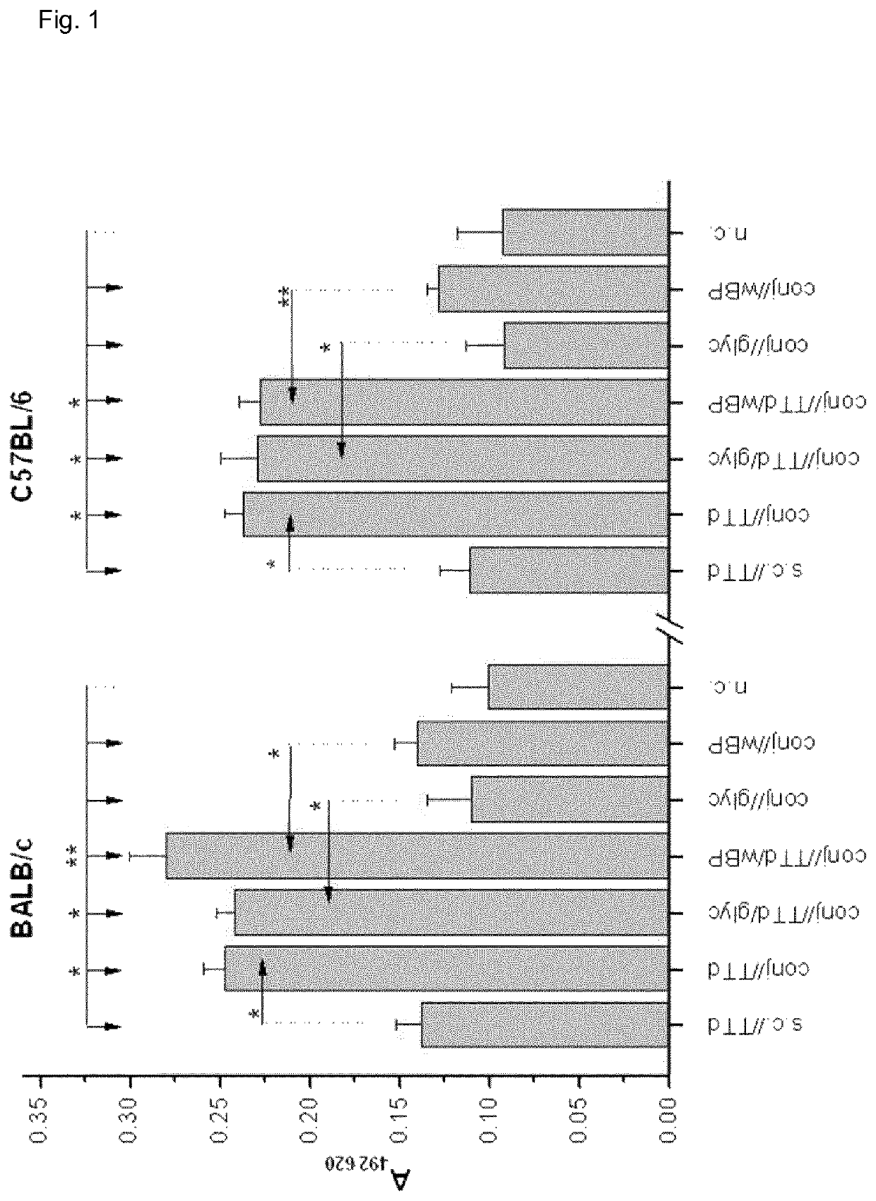
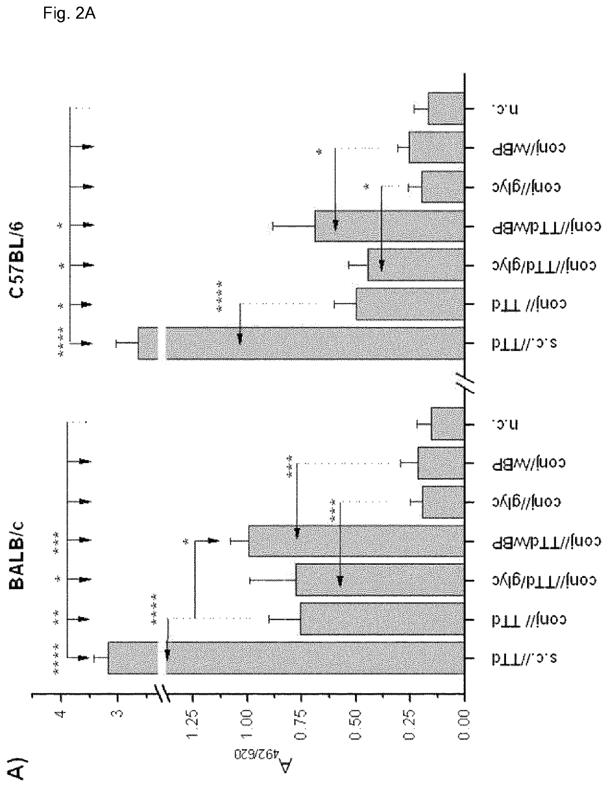
[0163]Eight-week-old BALB / c and C57BL / 6 female mice were used in the experiments. All experiments were approved by the “Ethics Committee for the Welfare of Experimental Animals” and by the committee section at the Institute of Virology, Vaccines and Sera—Torlak and conformed to the Serbian laws and European regulations on animal welfare (Approval No. 011-00-00510 / 2011-05 / 2).

[0164]Antigens, Adjuvants and Immunization and Bleeding Schedules

[0165]BALB / c and C57BL / 6 female mice were immunized via the conjunctiva (conj / / ) with TTd (Institute of Virology, Vaccines and Sera—Torlak, Belgrade, Serbia) as a model antigen (100 μg TTd / PBS per mouse in 10 μl (5 μl per eye) was applied to the conjunctiva), and 2% glycerol (glyc) and merthiolat-inactivated whole cell B. pertussis (wBP) (Institute of Virology, Vaccines and Sera—Torlak, Belgrade, Serbia) were used as Th1-promoting adjuvants. These adjuvants were chosen because glyc is commonly used in eye drops for human use...

example 2

Materials and Methods

Cell Culture

[0216]HCjE cells, kindly provided by Prof. Ilene Gipson (Schepens Eye Research Institute, HarvardMedical School, Boston), were maintained in keratinocyte serum-free medium (LifeTechnologies, Paisley, UK) at 37° C. / 5% CO2 and 95% humidity. The medium was changed every second day, and the cells were passaged at 70% confluence. Cells were harvested by trypsinisation (0.05% Trypsin / 0.02% EDTA in PBS, PAA Laboratories GmbH, Pasching, Austria) and seeded at a density of 30.000 cells / well in 6-well plates (Greiner Bio-One, Kremsmünster, Austria) for subsequent flow cytometric analysis and in 24-well imaging plates (PAA Laboratories GmbH, Pasching, Austria) for laser scanning microscopy.

Corpuscular Adjuvants of Bacterial Origin Labelled with Atto488 Dye

[0217]Lyophilised corpuscular adjuvants of bacterial origin were reconstituted in 0.1 M sodium bicarbonate buffer (pH 8.5, PAA Laboratories GmbH, Pasching, Austria) and incubated with Atto488 dye (2 mg / ml in D...

example 3

Experiments Concerning the Uptake of Corpuscular Adjuvants of Bacterial Origin by HCjE Cells

[0232]The influence of chitosan on the uptake of corpuscular adjuvants of bacterial origin by HCjE cells is tested.

Methods:

[0233]HCjE cells were seeded onto chamber slides at a density of 2×10⁸ cells / well and incubated at 37° C. overnight. 2×10⁸ of corpuscular adjuvants of bacterial origin labelled with ATTO-390 (Sigma-Aldrich, St. Louis, Mo.) dissolved in boric acid buffer (BAB), 0.05% chitosan-fluorescein isothiocyanate (FITC; Akina, USA) dissolved in BAB, and 2×10⁸ of labelled corpuscular adjuvants of bacterial origin dissolved in 0.05% labelled chitosan were added to separate wells containing HCjE cells for 30 minutes. The cells were washed with PBS, quenched with 0.4% trypan blue for 5 minutes and stained with CellMask plasma membrane stain (2.5 μg / ml; Molecular Probes, Inc., Eugene, Oreg.) for 5 minutes, both at 37° C. Then the cells were fixed with 4% PFA, mounted and examined by epifl...


PUM

PropertyMeasurementUnit
particle sizeaaaaaaaaaa
sizeaaaaaaaaaa
sizeaaaaaaaaaa

Abstract

The present invention relates generally to the field of ocular therapeutics and the development thereof for use in humans and animals including mammals and birds. More particularly, it relates to subunit vaccines that are effective against pathogens causing infections thereof for use in humans and animals including mammals and birds. The present invention specifically provides a novel vaccine formulation suitable for ocular immunization comprising a subunit vaccine antigen in an amount to provoke a protective immune response and at least two adjuvants of which one is corpuscular. It further provides a method for inducing a local and systemic immune response and methods for preventing recurrence of ocular infections and / or modulates the occurrence and / or severity of sequels.

Description

CROSS-REFERENCE TO RELATED APPLICATIONS[0001]This application is the U.S. national stage of International Patent Application No. PCT / EP2014 / 058390, filed on Apr. 24, 2014 and entitled NOVEL VACCINE FORMULATION FOR OCULAR IMMUNIZATION, which claims the benefit of priority under 35 U.S.C. § 119 from European Patent Application No. 13165103.6, filed Apr. 24, 2014. The disclosures of the foregoing applications are incorporated herein by reference in their entirety.FIELD OF THE INVENTION[0002]The present invention relates generally to the field of ocular therapeutics and the development thereof for use in subjects, including mammals, humans and birds. More particularly, it relates to subunit antigen vaccines that are effective against pathogens causing infections thereof for use in subjects.[0003]The present invention specifically provides a novel vaccine formulation suitable for ocular immunization of a subject comprising a subunit vaccine antigen in an amount to provoke a protective im...

Claims


Application Information

Patent Timeline
Patent Type & Authority Patents(United States)
IPC IPC(8): A61K39/08A61K39/39A61K9/00A61K47/06A61K31/7052A61K39/00
CPCA61K47/06A61K31/7052A61K9/0048A61K39/08A61K39/39A61K2039/6006A61K2039/54A61K2039/55594A61K2039/55583A61K2039/545A61K2039/6087
Inventor BARISANI-ASENBAUER, TALININIC-KANADA, ALEKSANDRA
Owner MEDIZINISCHE UNIVET WIEN
Why Eureka
Try Eureka