
Compositions and methods for platelet enriched fibrin constructs

a technology of fibrinogen and constructs, which is applied in the field of compositions and methods of platelet enriched fibrin constructs, can solve the problems of fibrinogen sealants, large handling difficulties of glues, and formation of fibrinogen sealants

Active Publication Date: 2020-12-01
DEPUY SYNTHES PROD INC
View PDF14 Cites 0 Cited by
  • Summary
  • Abstract
  • Description
  • Claims
  • Application Information

AI Technical Summary

Benefits of technology

The fibrin construct is dimensionally stable, allowing for secure delivery and adherence to non-horizontal surfaces, and effectively promotes wound healing by providing a sustained release of growth factors, enhancing tissue repair and regeneration.

Problems solved by technology

The fibrin sealants, formed by mixing a concentrated solution of fibrinogen with thrombin and calcium ions to produce fibrin, are impractical because the preparation method requires several hours and results in a crude clotting factor concentrate that is useful to manage hemostatically-deficient patients, but is not practical for harvesting fibrinogen from small volumes of blood.
Fibrin glues mimic the last step of the coagulation cascade and are usually commercialized as kits comprising components to form a three-dimensional network commonly called “Fibrin Gel.” However, an important and well known disadvantage of the known fibrin glue preparations resides in the water-like fluidity of the components, which leads to considerable handling difficulties of the glues.
The basic problem with a low viscosity glue still remains.
Multiple problems exist with the present tissue sealants.
First, a non-viscous or low viscosity glue is unsuitable for use on non-horizontal surfaces since it will run off before setting.
Second, there is a definite risk of a non-viscous or low vicosity glue running off to sites where it is unwanted and where it might cause complications.
This is particularly the case in vascular surgery since the fluid glue may reach inside the vessels before it sets and thereby cause thromboembolic complications.
An instantaneously setting fibrin glue (containing a high concentration of thrombin), on the other hand, cannot be used where the parts to be sealed require subsequent adaptation.
Moreover, the present solutions do little to expedite wound healing.
However, delivery vehicles for growth factors are not always reliable or able to remain in the desired location for a sufficient period of time.
Moreover, formulation and delivery of multiple growth factors, which are often needed for improved healing, are not practical due to cost and availability.

Method used


Image

  • Compositions and methods for platelet enriched fibrin constructs
  • Compositions and methods for platelet enriched fibrin constructs
  • Compositions and methods for platelet enriched fibrin constructs

Examples


example 1

on of Platelet Enriched Fibrin Construct (PEFC)

[0131]Soft Spin Preparations:

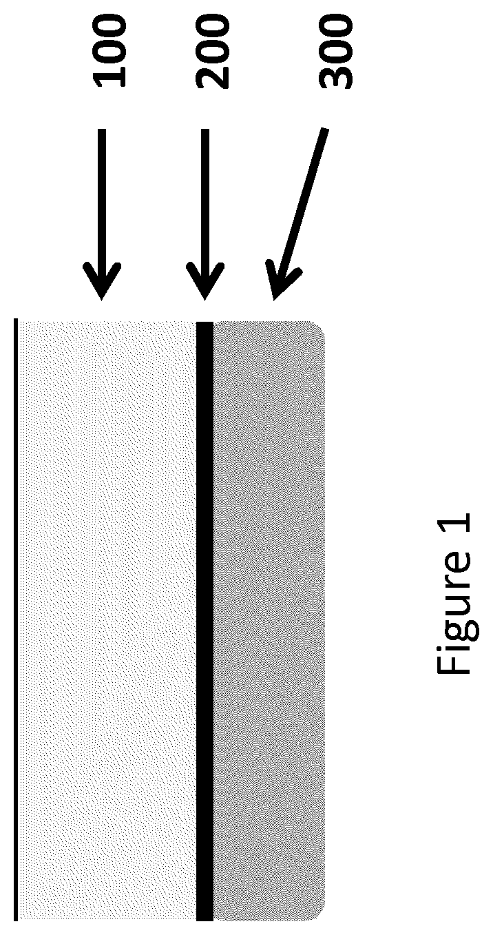
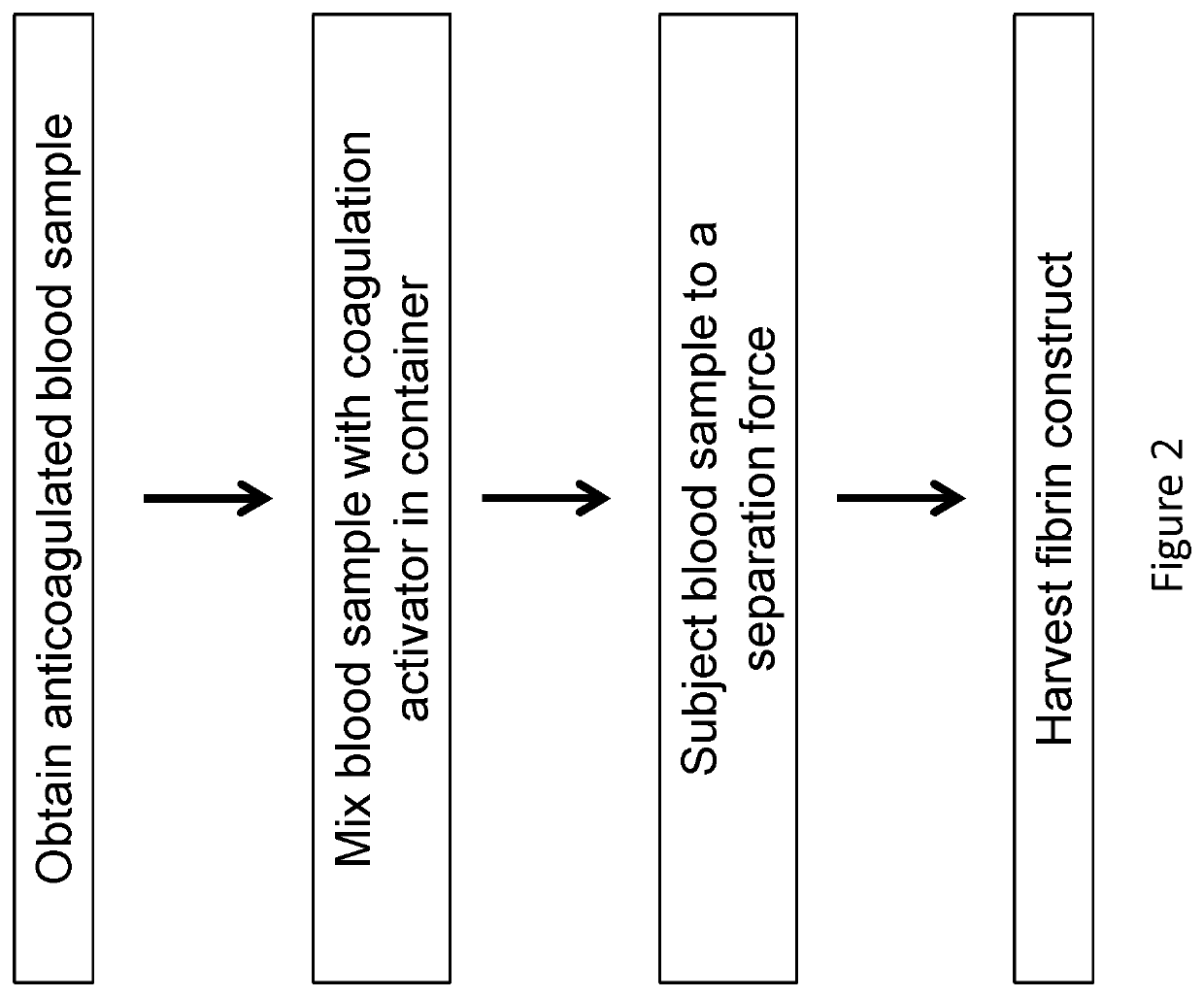
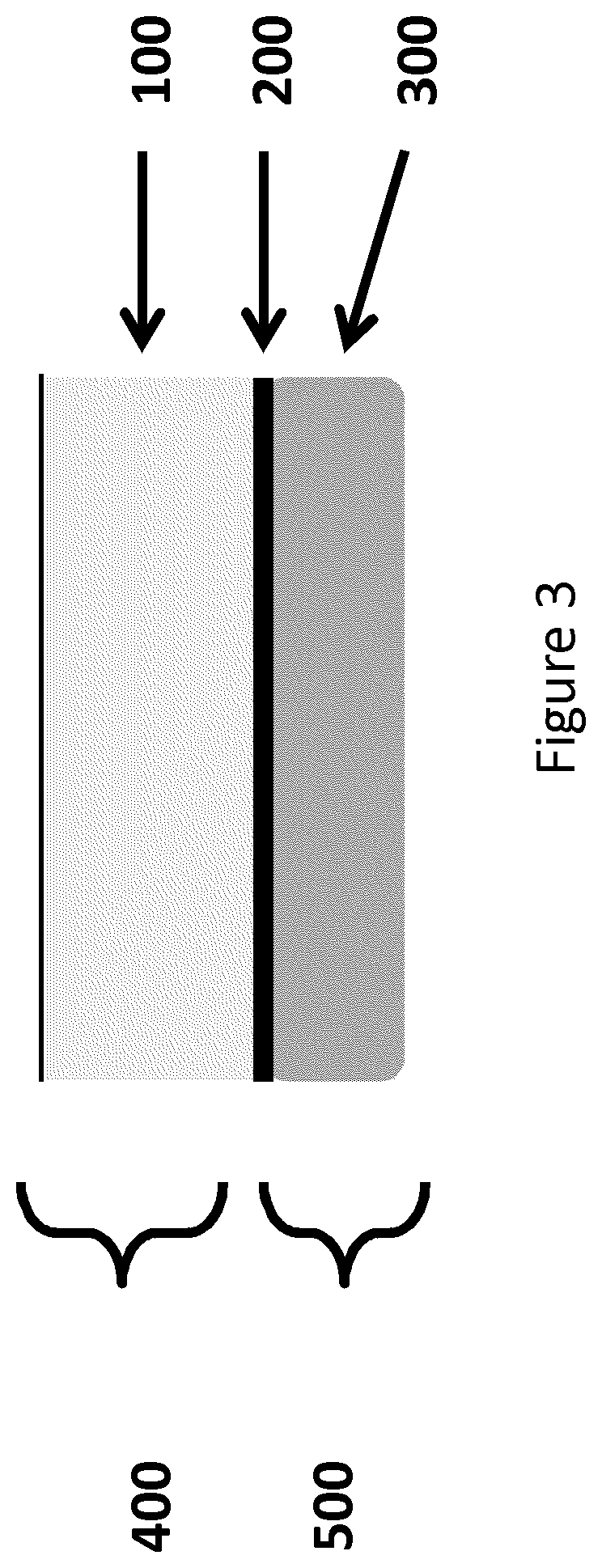
[0132]Bovine blood samples of 6.8 ml were collected and exposed to 1.2 ml of anti-coagulant, ACD-A (15% by volume in final mixture). The blood samples were transferred to borosilicate glass tubes with 180 μl of 1M CaCl2 solution. The samples were inverted 10 times and centrifuged at 2000×g for 10 mins. During centrifugation, the blood sample clotted and separated into distinct layers: a plasma layer (PLASMA), a fibrin plug layer (CLOT) and a blood cell layer (RBC). The images of FIG. 9 sequentially illustrate this procedure.

[0133]The blood cell layer can be physically removed by peeling the cap of blood cells, as shown in FIGS. 4A and 4B, from the fibrin layer, resulting in a construct comprising platelet / white blood cells, as shown in FIG. 4C. Also, FIG. 4D illustrates the blood cell cap removed from the fibrin layer. Most of the red blood cells can further be removed by blotting, scraping, etc.

[0134]Double...

example 2

Properties of the Platelet Enriched Fibrin Construct

[0145]Physical property tests of the fibrin constructs were conducted with constructs prepared with bovine blood collected with ACD-A and shipped the previous day. A total of 18 samples were produced consecutively, with the maximum of 6 samples in centrifuge at a time for 15 minutes at 4500 RPM (2820 g) in capped borosilicate test tubes (with 180 μl of 1M CaCl2 solution). Upon the completion of the spin, the test tubes were immediately removed and placed vertically in holders. The clots, which were ˜14 mm in diameter, were extracted one at a time using forceps; excess proximal protein was removed, excess fluid was initially removed with gauze, and then the construct was trimmed to reduce thickness to ˜1 mm after a moderate amount of fluids was removed. The constructs were stored in DPBS solution until all samples were prepared and the DPBS fluids were removed from the constructs with light compression with gauze before mechanical t...

example 3

al Properties of the Platelet Enriched Fibrin Construct

[0158]Histology:

[0159]Human PEFC clots were generated according to the procedure of Example 1. Clots were gently blotted on kimwipes to remove excess fluid, cut into 3 sections and mounted in OCT compound on dry ice so that the cut edge created the plane for sectioning. Frozen cassettes were then sectioned in a cryostat ultra microtome at a thickness of approximately 5 microns. Tissues were immediately fixed in 100% methanol for 5 minutes at room temperature after mounting on glass slides. Slides were rinsed in phosphate buffered saline and then subjected to Wright-Giemsa staining following a standard whole blood smear staining protocol. Photomicrographs were obtained from the same field of view at increasing magnifications, as indicated in FIG. 15, using a Zeiss microscope outfitted with a digital color CCD camera.

[0160]FIG. 15 shows the stained sections of the PEFC at 50×, 100× and 200×. The upper non-stained portion is the fi...


PUM

PropertyMeasurementUnit
concentrationaaaaaaaaaa

Abstract

Compositions and methods are provided for tissue constructs that promote wound healing. The composition comprises a dimensionally stable fibrin construct for local administration to a wound site or region. In one embodiment, the fibrin construct is a wound healing composition, including components that promote wound healing, such as platelets, growth factors, white blood cells and fibrin clots. In another embodiment, the tissue treatment composition includes (i) aggregated fibrin, (ii) blood cells, and (iii) optionally, growth factors and / or other proteins.

Description

FIELD OF THE INVENTION[0001]The present invention relates generally to compositions and methods of platelet enriched fibrin constructs for delivery.BACKGROUND OF THE INVENTION[0002]Wound healing, or wound repair, is an intricate process in which the skin (or another organ-tissue) repairs itself after injury. In normal skin, the epidermis and dermis exists in a steady-state equilibrium, forming a protective barrier against the external environment. Once the protective barrier is broken, the normal (physiologic) process of wound healing is immediately set in motion. The classic model of wound healing is divided into three or four sequential, yet overlapping phases: (1) hemostasis, (2) inflammatory, (3) proliferative and (4) remodeling. Upon injury to the skin, a set of complex biochemical events takes place in a closely orchestrated cascade to repair the damage. Within minutes post-injury, platelets (thrombocytes) aggregate at the injury site to form a fibrin clot. This clot acts to c...

Claims


Application Information

Patent Timeline
Patent Type & Authority Patents(United States)
IPC IPC(8): A61K35/14A61K38/18A61K35/17A61L27/38A61K38/55A61K38/36A61L15/32A61L27/54A61K35/19A61L27/22
CPCA61K35/19A61K38/363A61K35/14A61L27/225A61L15/32A61K38/1808A61K38/1866A61K38/18A61K38/1825A61L27/54A61K38/1841A61K38/1875A61L27/38A61K35/17A61K38/55A61K2300/00A61L2300/414A61P43/00
Inventor SENGUN, MEHMET Z.PARRISH, WILLIAMWHITTAKER, GREGORY R.FIFOLT, DOUGLAS A.STORY, BROOKS J.
Owner DEPUY SYNTHES PROD INC
Learn More