Liver-mimetic device and method for simulation of hepatic function using such device

a liver-mimetic device and liver-mimetic technology, which is applied in the field of liver-mimetic devices and methods for simulation of hepatic functions using such devices, can solve the problems of difficult to translate human studies, costing the traditional animal model, and affecting the success of drug development, and achieves rapid and scalable fabrication of highly-specified biomimetic structures

Active Publication Date: 2021-03-23
RGT UNIV OF CALIFORNIA
View PDF5 Cites 1 Cited by
  • Summary
  • Abstract
  • Description
  • Claims
  • Application Information

AI Technical Summary

Benefits of technology

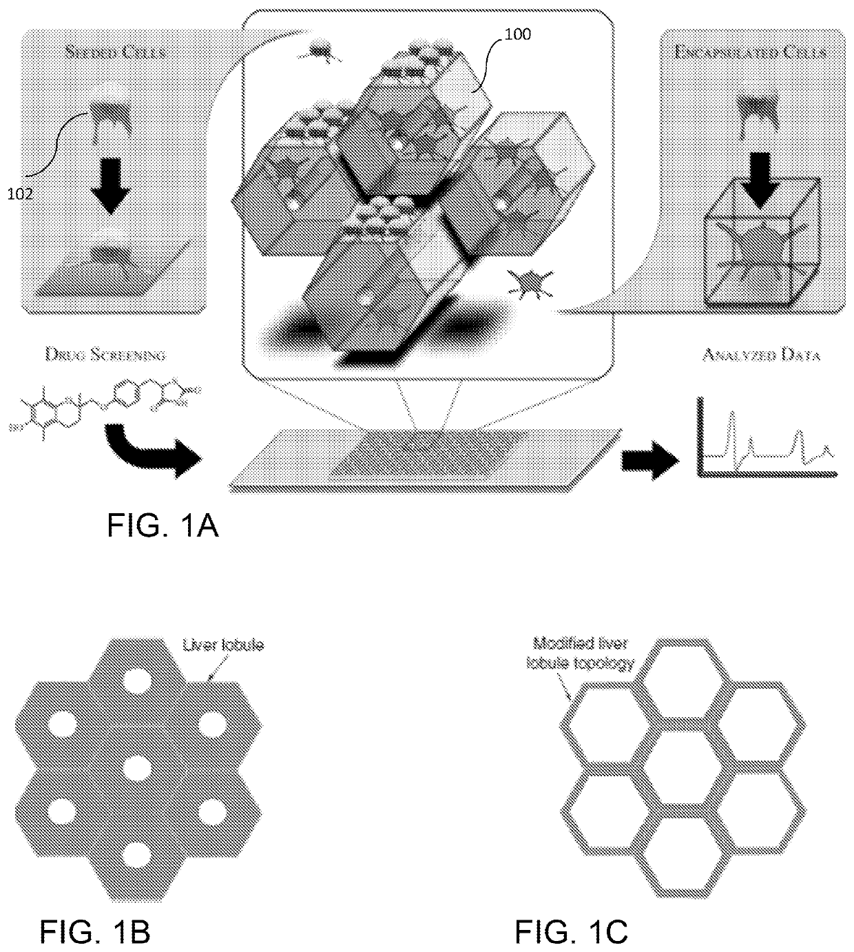
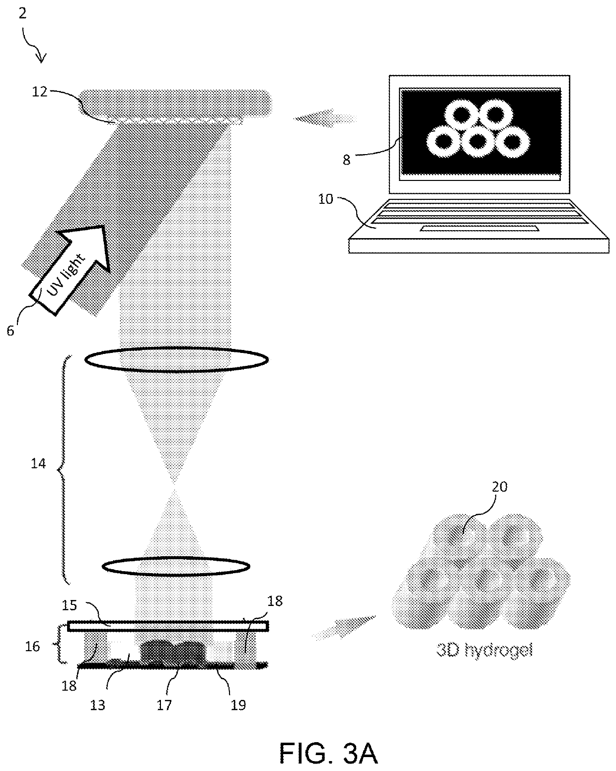
[0010]According to embodiments of the invention, dynamic optical projection stereolithography (“DOPsL”) is used for rapid, scalable fabrication of highly-specified biomimetic structures. In particular, a 3D liver-mimetic structure can be fabricated using DOPsL to closely mimic hepatic micro-architecture and function by encapsulating particles that are capable of performing one or more liver function. These particles can be cells or polymer nanoparticles.
[0011]In some embodiments, a functional in vitro micro-liver model can be produced via encapsulation of hepatic progenitor cells (HPCs) derived from human iPSCs using dynamic optical projection stereolithography (“DOPsL”). This advanced model consists of co-culture of HPCs and supportive cells (endothelial cells and mesenchymal stem cells) within a biomimetic scaffold closely mimicking hepatic micro-architecture. This patient specific liver-on-a chip model can be explored as a reliable and cost-efficient in vitro platform to facilitate drug metabolism studies, preclinical drug screening, and fundamental hepatology research.
[0014]The DOPsL technology utilizes a digital mirror array device (DMD) to generate dynamic photomasks that can be translated into a 3D complex structure through layer-by-layer photopolymerization of biomaterials. The DOPsL technology has demonstrated considerable versatility in fabricating 3D complex geometries for functional devices and even artificial tissues, providing for the rapid and scalable fabrication of highly-specified biomimetic structures. This approach provides capability in terms of speed and scalability that cannot be well-achieved using existing technologies that employ raster-based printing approaches or soft lithography techniques. Additionally, as the DOPsL system can accept any set of high detail images, this platform provides a method for generating models with patient specificity not only in terms of cell type but also whole tissue morphology. Furthermore, the flexibility of the platform allows for the modular addition and subsequent decoupling of various components of a complex 3D construct, providing a means to determine the individual contributions of material type, co-culture populations, spatial cell arrangements, and biomimetic geometry towards recapitulating native liver physiology. Using the DOPsL approach, appropriate liver-on-a-chip models can be fabricated for various application contexts from fundamental hepatic functionality to high throughput screening for pharmacotoxicity.
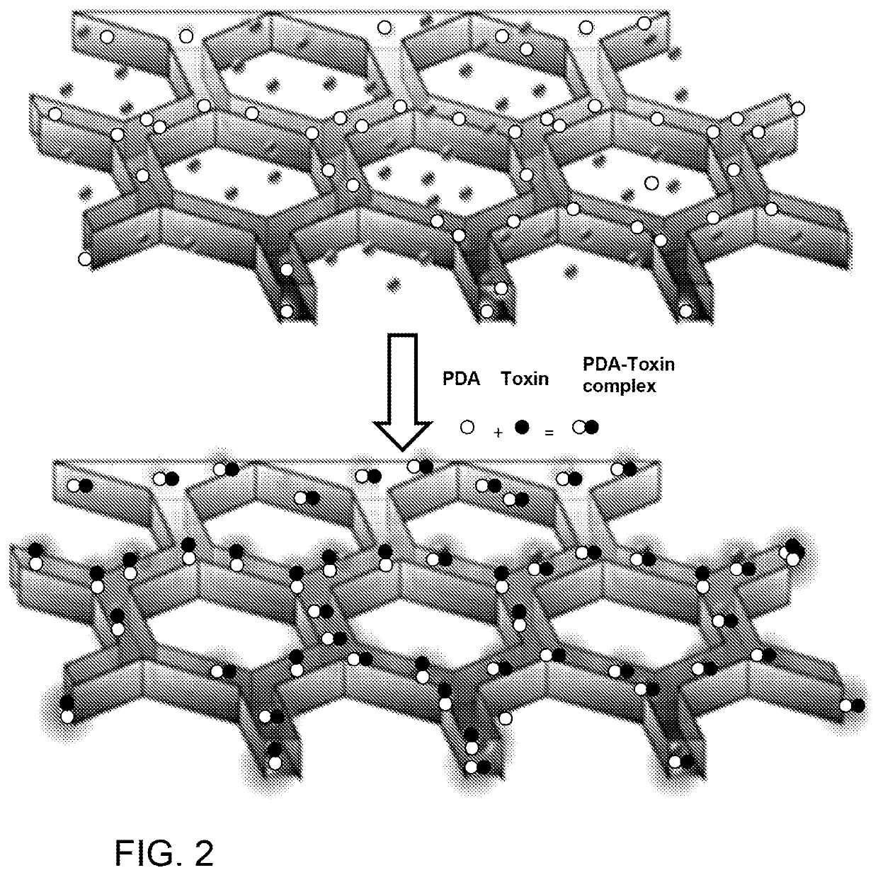
[0015]Utilizing biofabrication, a 3D matrix can be formed with a modified liver lobule configuration to encapsulate nanoparticles and / or cells that possess hepatic function. This liver-on-a-chip in vitro model provides a tool for a variety of applications. In addition to detoxification, the device may also serve as a versatile instrument for cost-effective and reliable early-stage drug targeting and preclinical drug screening. Such a device has the potential of markedly reducing the high cost of drug development, now a major economical roadblock to sustained progress in healthcare.

Problems solved by technology

Unfortunately, the dramatic discrepancy between available donors vs. patients on the waiting lists highlights the critical need for functional liver replacements.
Conventional animal models are costly, often unreliable, and difficult to translate to human studies due to the species-specific variations in hepatocellular functions.
The resulting financial consequences of a drug failing at the clinical stage can be catastrophic to the drug makers.
While several human liver models, such as liver slices, microsomes, cell lines and primary hepatocytes, are currently in use, these models are still limited in terms of fully representing the dynamic cellular responses of healthy liver tissue.
Nonetheless, cell sourcing remains a significant challenge for both in vivo and in vitro liver models.
Most tissue engineered liver models consist of suboptimal cell sources, such as primary hepatocytes and hepatic cell lines isolated from liver tumors (e.g. HepG2), which make them less practical for clinical applications.
However, none of these models exhibit similar micro-architecture compared to native liver tissue.
Conventional cultures of primary hepatocytes have served as the gold standard for in vitro applications, however, these primary cells lack robust proliferative capacity and are prone to de-differentiate fairly quickly.
As a result, their functional activity can only be maintained for a relatively short time frame (i.e., 24-72 hours).
Furthermore, human primary hepatocytes are difficult to obtain on a consistent basis.
Improvements in cell sourcing notwithstanding, the development of fully functional liver constructs has been limited due to the challenge of fully recapitulating the native physical structure of hepatic tissue.
However, very limited progress has been achieved in fully mimicking the native architecture and organization of liver in both composition and form, partially due to the lack of appropriate biofabrication techniques.
Conventional detoxification platforms such as antisera, monoclonal antibodies and small-molecule inhibitors are unable to completely neutralize toxins because of their limited ability to block the entire PFT molecule.
Although patients suffering from poisoning may benefit from these strategies, intravenous administration of nanoparticles can cause nanoparticle-toxin accumulation in the liver, leading to the risk of secondary poisoning, which is particularly detrimental in liver-failure patients.

Method used


Image

  • Liver-mimetic device and method for simulation of hepatic function using such device
  • Liver-mimetic device and method for simulation of hepatic function using such device
  • Liver-mimetic device and method for simulation of hepatic function using such device

Examples


example 1

Evaluation of Hepatic Maturation and Function

[0060]The maturation and function of HPCs in the 3D scaffold can be systematically evaluated by commonly-used assays. The gene expression of ALB, HNF 4α, G6D, and MRP 2 are assessed by real time PCR at days 3, 7, 21 and 30. Albumin secretion and urea synthesis may also be monitored at the same time points to further confirm hepatic maturation. ELISA is used to quantify the albumin secretion at each time point. Commercially available kits (Sigma-Aldrich, Inc.) can be used to measure the urea concentration in the culture at the same time points.

[0061]After confirming hepatic maturation, further tests on hepatic function may be performed. Gluconeogenesis can be probed using periodic-acid Schiff (PAS) staining. Cytochrome P450 enzymatic activity, which plays an important role in detoxification processes, can be determined via P450-Glo™ CYP450 Assays (Promega). Formation of bile canaliculi is characterized as well at day 7, 21 and 30. After th...

example 2

Optimization of Co-culture Conditions

[0063]The liver consists of multiple cell types, such as hepatocytes, endothelial cells, and fibroblasts, working together to provide hepatic functionality. For hepatic cultures in vitro, it has been widely recognized that co-cultures with other supportive cells, e.g., endothelial cells, fibroblasts, and MSCs, can significantly stabilize hepatic phenotype and function. In order to achieve the most successful co-culture system, optimization of culture conditions (e.g., combination of culture media) should be performed both in 2D and 3D contexts prior to incorporating cells within the complex 3D scaffold. In prior studies and preliminary tests, co-cultures of MSCs and endothelial cells with hepatocytes are well supported in hepatic media.

[0064]Vascularization is vitally important for tissues with highly active metabolism, such as the heart and liver due to their intensive demand of oxygen and nutrient supply as well as waste disposal. By incorporat...


PUM

PropertyMeasurementUnit
timeaaaaaaaaaa
concentrationsaaaaaaaaaa

Abstract

A liver-mimetic device and method include a 3D polymer scaffold having a matrix of liver-like lobules with hepatic-functioning particles encapsulated within the lobules. In some embodiments, each liver-like lobule is hexagonal in structure and the matrix is in a honeycomb arrangement. In some embodiments, the hepatic-functioning particles are hepatic progenitor cells. In other embodiments, the hepatic-functioning particles are polymer nanoparticles adapted to capture pore-forming toxins.

Description

RELATED APPLICATIONS[0001]This application is a 371 national stage filing of International Application No. PCT / US2014 / 040946, filed Jun. 4, 2014, which claims the benefit of the priority of U.S. Provisional Application No. 61 / 831,100, filed Jun. 4, 2013 and U.S. Provisional Application No. 61 / 927,906, filed Jan. 15, 2014.GOVERNMENT RIGHTS[0002]This invention was made with government support under Grant EB017876 awarded by the National Institutes of Health. The government has certain rights in the invention.FIELD OF THE INVENTION[0003]The present invention relates to a liver-mimetic device fabricated using a 3D printed hydrogel matrix for encapsulating particles capable of performing hepatic functions and a method for simulation of hepatic function with a liver-mimetic device.BACKGROUND[0004]In the U.S., liver associated diseases are major contributors to morbidity and mortality. Approximately 40,000 people in the U.S. die each year from acute or chronic liver diseases. Organ transpl...

Claims


Application Information

Patent Timeline
Patent Type & Authority Patents(United States)
IPC IPC(8): C12N5/071
CPCC12N5/0671C12N2506/45C12N2533/30C12N2533/54C12N2533/80
Inventor QU, XINGOU, MALINGZHU, WEICHEN, SHAOCHEN
Owner RGT UNIV OF CALIFORNIA
Why Eureka
Try Eureka