
Anti-TGF-β induction of bone cell function and bone growth

a technology of anti-tgf- and bone cell function, which is applied in the field of molecular biology and medicine, can solve the problems of hardly the best approach to therapy, structural instability, and failure to repair defects, and achieve the effect of increasing bone mass and/or volum

Active Publication Date: 2021-12-07
VANDERBILT UNIV
View PDF13 Cites 0 Cited by
  • Summary
  • Abstract
  • Description
  • Claims
  • Application Information

AI Technical Summary

Benefits of technology

The anti-TGF-β antibody treatment significantly increases bone volume, density, and strength by enhancing osteoblast numbers and reducing osteoclast activity, leading to improved bone quality and reduced fracture risk, as demonstrated in preclinical studies.

Problems solved by technology

Osteoporosis represents a large and rapidly growing health care problem with an unmet medical need for therapies that stimulate bone formation.
Most current drugs for osteoporosis retard bone degradation but do not stimulate bone formation to replace already lost bone.
These complications are recognized and measures are usually taken to avoid them, but these are hardly the best approach to therapy.
Yet another bone-related health issues is bone reconstruction and, specifically, the ability to reconstruct defects in bone tissue that result from traumatic injury, as a consequence of cancer or cancer surgery, as a result of a birth defect, or as a result of aging.
However, experience has shown that a lack of viable bone bridging the defect can result in exposure of the appliance, infection, structural instability and, ultimately, failure to repair the defect.
Autologous bone grafts are another possibility to deal with bone injury, but they have several demonstrated disadvantages in that they must be harvested from a donor site such as iliac crest or rib, they usually provide insufficient bone to completely fill the defect, and the bone that does form is sometimes prone to infection and resorption.
Partially purified xenogeneic preparations are not practical for clinical use because microgram quantities are purified from kilograms of bovine bone, making large scale commercial production both costly and impractical.
However, these techniques are time consuming, have been shown to produce a great deal of morbidity, and can only be used by specially trained individuals.
A number of cancers metastasize to bone and can result in bone weakening, and some are even associated with bone destruction and bone loss, such as breast, lung, thyroid, kidney and prostate cancer.

Method used


Image

  • Anti-TGF-β induction of bone cell function and bone growth
  • Anti-TGF-β induction of bone cell function and bone growth
  • Anti-TGF-β induction of bone cell function and bone growth

Examples


example 1

Materials & Methods

[0140]Antibodies.

[0141]The 1D11 antibody was generated by Genzyme Corporation (Framingham). Control antibody (13C4) consistent of an identical IgG complex lacking any TGF-β binding capabilities.

[0142]Treatment Regimen.

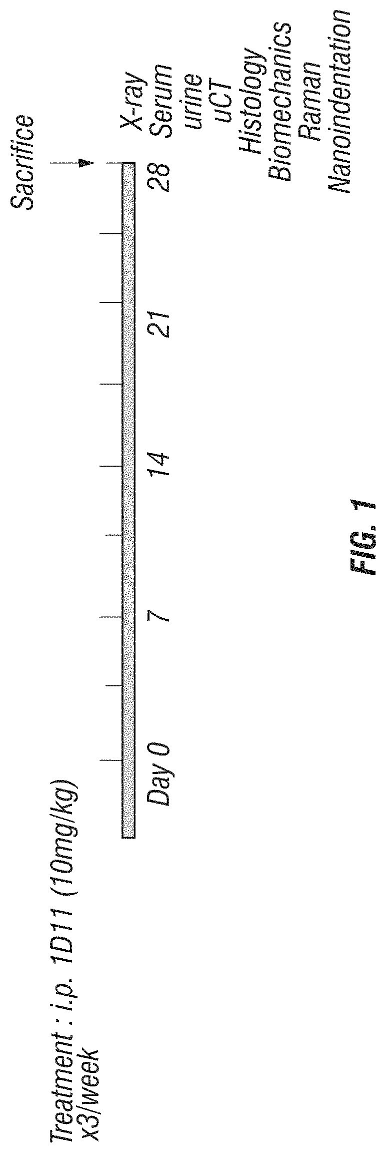
[0143]Normal 13-week old male C57Bl / 6 mice (Harlan) (n=5) were treated with 10 mg / kg / ×3 week of 1D11 or control antibody. Each reagent was administered by sterile intra-peritoneal injection over a 4 week time period (FIG. 1). All animal procedures were conducted in accordance with IACUC protocols approved by Vanderbilt University Medical Center.

[0144]Imaging.

[0145]Tibia and femure were analyzed by μCT scanning (μCT40, Scanco) at an isotropic voxel size of 12 μm (55Kv). After the growth plate was identified in each scan set, the metaphyseal region 200 μm below this area was scanned and analyzed for alterations in trabecular bone parameters (Threshold 280).

[0146]Histology and Histomorphometry.

[0147]Lumbar vertebral bodies (L3-5) and long bones were col...

example 2

Results

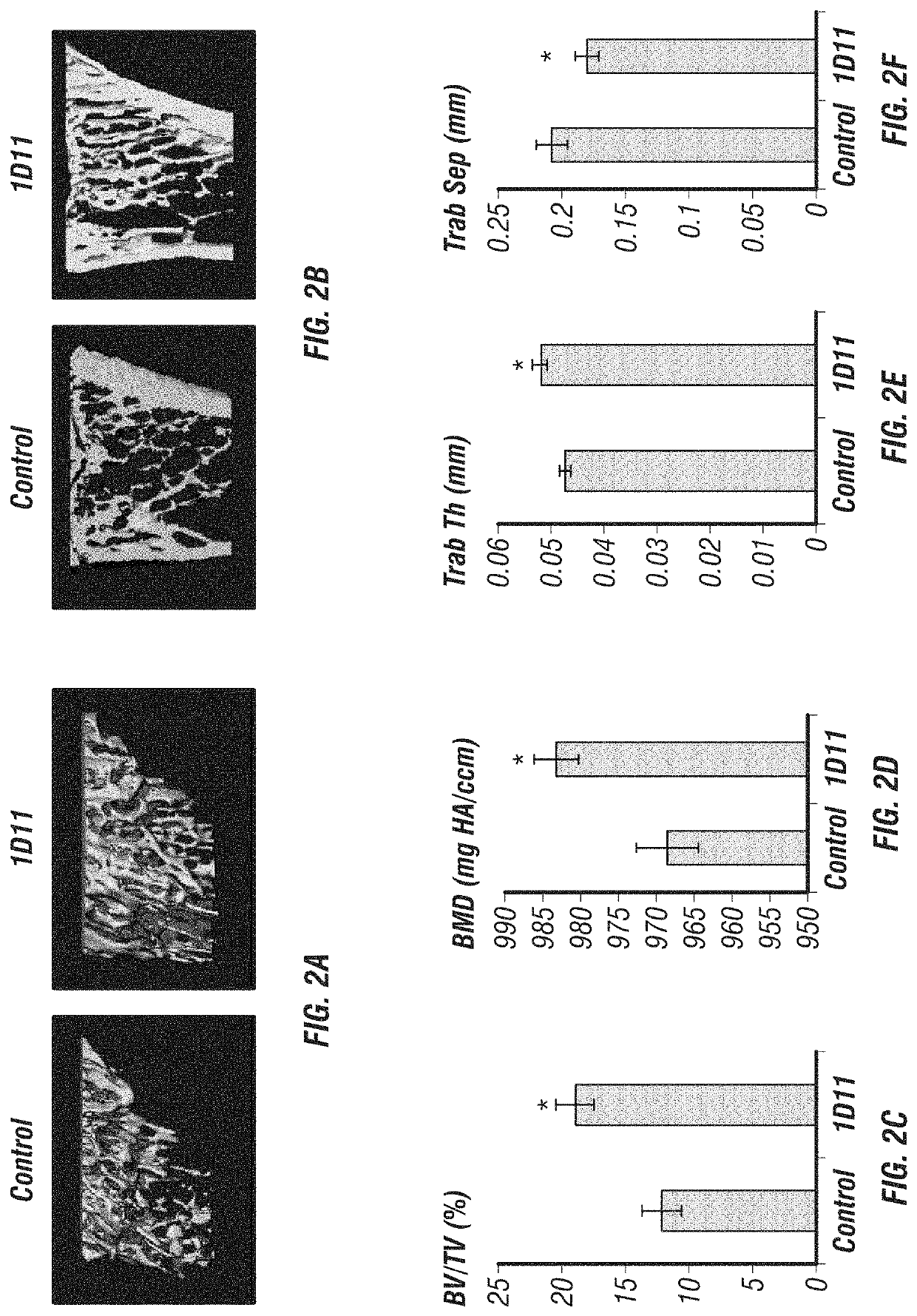
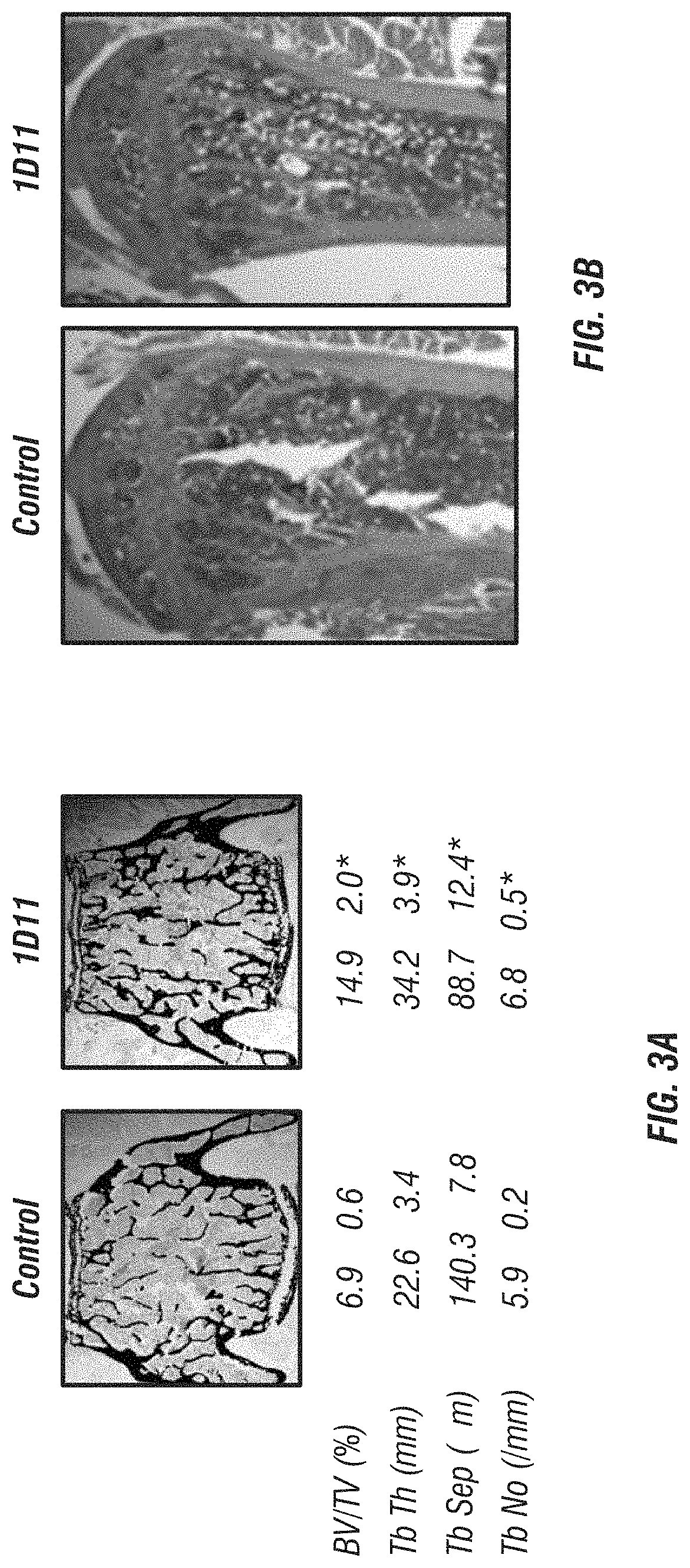
[0162]Inhibition of TGF-β by 1D11 antibody treatment, outlined in FIG. 1, significantly increased long bone volume compared to controls (FIGS. 2A-B). Trabecular bone at the tibial metaphysis analyzed by μCT scanning showed a dramatic increase in overall BV / TV (FIG. 2C), bone mineral density (BMD) (FIG. 2D), trabecular thickness (FIG. 2F) and decreased trabecular separation (FIG. 2F) in animals treated with 1D11 compared to control-treated mice. Histomorphometric analysis of undecalcified sections from lumbar vertebra supported μCT analysis of long bones. 1D11-mediated TGF-β inhibition led to a 54% increase in trabecular BV / TV. This increase in bone was accompanied by greater trabecular number, decreased trabecular separation and increased trabecular thickness (FIGS. 3A-B).

[0163]An analysis of bone cell distribution in TRAP-stained vertebral sections showed significantly reduced osteoclast numbers and surface area following 1D11 treatment (FIGS. 4A-C). In contrast, elevated os...

example 3

Discussion

[0167]This study investigates the use of a TGF-β neutralizing antibody as an anabolic bone agent and highlights the potential of TGF-β inhibition as a mechanism to increase bone mass. The inventors employed standard, accepted techniques along with emerging technologies to thoroughly analyze bone volume, density, strength and composition. Together, these studies demonstrate that drugs aimed at blocking the TGF-β signaling pathway have the capacity to positively regulate osteoblast numbers while simultaneously decreasing the amount of active osteoclasts in the marrow. This results in a profound increase in bone volume and quality, similar to that seen in PTH-treated rodent studies (Dempster et al., 1993).

[0168]There is a considerable need for more efficacious bone anabolic agents. Currently, the major therapeutic approach to excessive bone loss is through the use of anti-resorptives such as bisphosphonates. While these agents are certainly capable of repressing further bone ...


PUM


Abstract

The invention regards the modulation of TGF-β activity by administering to a subject an antibody that binds to TGF-β, thereby increasing bone growth, bone formation, bone mass and bone strength. The antibody acts to increase osteoblast number and function while at the same time decreasing osteoclast number and function. Such drugs are useful in the treatment of diseases or disorders such as osteoporosis, Paget's disease, metastatic bone cancer, myeloma bone disease, bone fractures, etc.

Description

[0001]This present application is a continuation of U.S. application Ser. No. 13 / 266,082, filed Jun. 25, 2012, now abandoned, which is a national phase application under 35 U.S.C. § 371 of International Patent Application No. PCT / US2010 / 032373, filed Apr. 26, 2010, which claims benefit of priority to U.S. Provisional Application No. 61 / 172,539, filed Apr. 24, 2009, the entire contents of each of which being hereby incorporated by reference.[0002]Monoclonal antibody 1D11 (IgG1) refers to the mAb produced by a clone of the murine hybridoma cell line 1D11. Samples of this cell line were deposited on Oct. 7, 1988 with the American Type Culture Collection (ATCC), 12301 Parklawn Drive, Rockville, Md. Cell line 1D11.16 and assigned ATCC HB9849, the publicly availability of which is guaranteed for thirty years after the issue date of this patent.BACKGROUND OF THE INVENTIONI. Field of the Invention[0003]The present invention relates to the fields of molecular biology and medicine. More parti...

Claims


Application Information

Patent Timeline
Patent Type & Authority Patents(United States)
IPC IPC(8): A61K39/395C07K16/22A61P19/08A61P19/10C07K16/24A61K39/00
CPCC07K16/24A61K39/3955A61P19/08A61P19/10C07K16/22A61K2039/505A61K2039/545C07K2317/76A61P19/00A61P35/00
Inventor EDWARDS, JAMES R.MUNDY, GREGORY R.
Owner VANDERBILT UNIV
Learn More