Use of heparinase to decrease inflammatory responses

a technology of heparin and inflammatory response, which is applied in the direction of antibodies, peptide/protein ingredients, lyases, etc., can solve the problems of affecting the afflicted individual, destroying viable cells and tissues, and causing harm to the afflicted individual, so as to reduce the side effects of treatment, inhibit leukocyte rolling and chemokine gradient formation, and reduce the effect of inflammatory respons

Inactive Publication Date: 2001-07-05
BIOMARIN PHARMA INC +1
View PDF0 Cites 37 Cited by
  • Summary
  • Abstract
  • Description
  • Claims
  • Application Information

AI Technical Summary

Benefits of technology

0053] Fusion proteins incorporating heparinase enzyme fused to proteins with specific binding properties can be created by recombinant molecular biology techniques. By choosing an appropriate binding protein, heparinase activity can be targeted to specific sites, in vivo. ICAM-1 has been shown to be preferentially expressed on the surface of activated endothelial cells (Dustin, et al., J. Immunol., 137:245-254, 1986). As examples of fusion proteins; an antibody, Fab fragment or variable region, specific for ICAM-1, VCAM-1 or P-selectin, when fused to heparinase enzyme or an active portion thereof, localizes heparinase activity near the luminal and abluminal surfaces of activated endothelium. Heparin and heparan sulfate moieties are removed in this area, causing breakdown of the chemokine gradient produced by the endothelium. As other examples, fusion of heparinase enzyme, or an active portion thereof, to the I-domain of LFA-1 or Mac-1 (both bind to ICAM-1) targets activated endothelium for removal of heparin and heparan sulfate, inhibiting leukocyte rolling and chemokine gradient formation. Receptors for cytokines such as IL-1b, are up-regulated on activated endothelium and provide another target for binding of fusion proteins. By fusing IL-1b, or the receptor binding domain of IL-1b to heparinase targeting can also be achieved. The fusion proteins can decrease inflammatory responses at lower blood concentrations than is required for comparable decreases using unfused heparinase. In addition, other cells in the vascular system will be less affected by the enzyme activity of the fusion protein, reducing possible side effects of treatments.
0054] Heparinase fusion proteins created by genetic engineering retain the binding and catalytic properties of heparinase and of the protein to which it is fused. Three heparinases have been purified to homogeneity from Flavobacterium heparinum, and have been produced in a recombinant form in Escherichia coli. Fusion proteins consisting of heparinase enzyme combined with binding domains from antibodies or adhesion molecules can be produced with a gene fusion in a recombinant host, while retaining the functionalities of binding and the enzymatic activity of the separate proteins. These molecules can also be purified to homogeneity by procedures normally used for purification of the individual parts of the fusion protein (e.g. affinity chromatography, heparinase purification protocols). Unlike the natural heparinase purified from Flavobacterium heparinum, the recombinant enzyme may not contain amino-terminal pyroglutamate or carbohydrate moieties. All recombinant heparinase may contain deletions, additional and / or altered amino acids, which modify the enzymatic activity of the natural enzyme or the functioning of the binding domains. Heparinase and fusion heparinase can be stabilized for in vivo use, by complexing with polyethylene glycol, cross linking agents, and by microencapsulation.

Problems solved by technology

While most localized inflammatory responses are beneficial, harmful inflammatory responses can occur.
Many harmful inflammatory responses also involve accumulation of leukocytes within a tissue.
This accumulation results in the destruction of viable cells and tissue.
In addition to damaging tissue, these responses are detrimental to, or debilitating for, the afflicted individual.
Because of difficulty in distinguishing regions of heparin and heparan sulfate on the same carbohydrate chain, little data exists on the binding preference of chemokines for either heparin or heparan sulfate moieties.
The antibodies interfere with the function of the adhesion molecules and block or reduce leukocyte recruitment.
The presence of soluble chemokine can interfere with adhesion and migration along a bound chemokine gradient.

Method used


Image

  • Use of heparinase to decrease inflammatory responses
  • Use of heparinase to decrease inflammatory responses
  • Use of heparinase to decrease inflammatory responses

Examples


example 1

of Endothelial Cells with Heparinase to Release Heparin / Heparan Sulfate

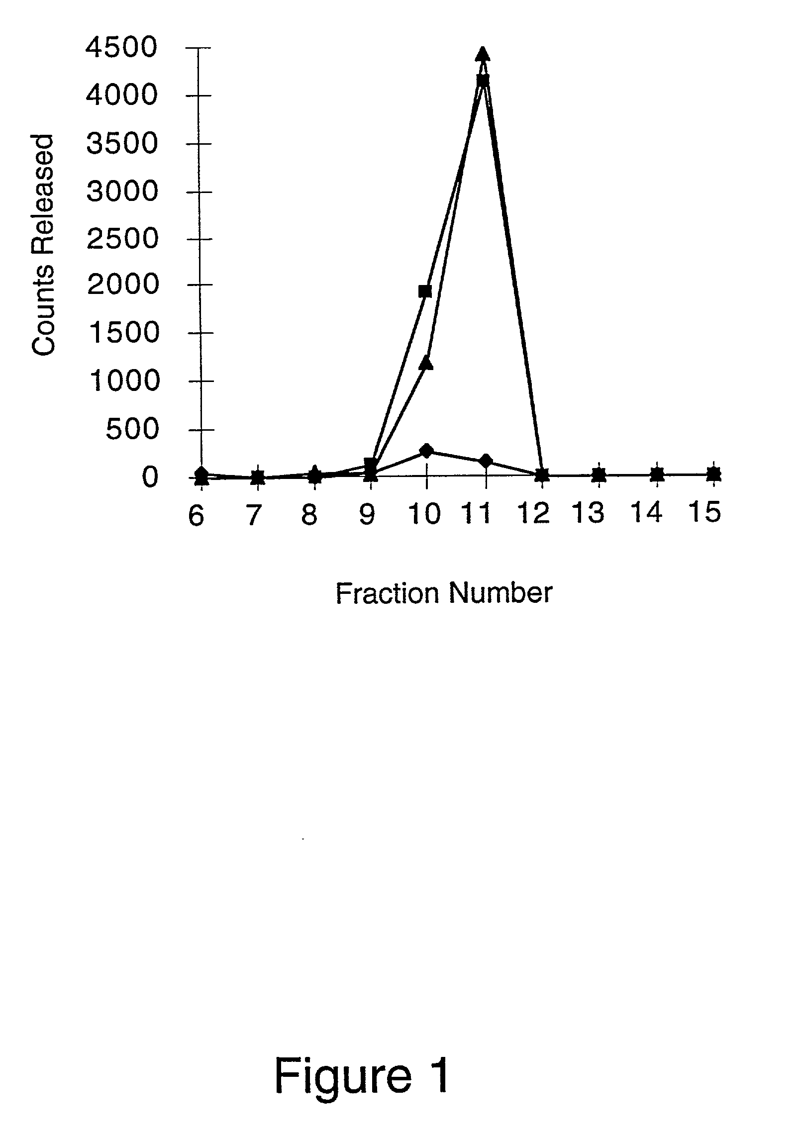
[0068] Heparinase alters the cell surface and basement membrane by cleaving the heparin and heparan sulfate moieties from the cell surface and extracellular matrix proteoglycans. Removal of these glycosaminoglycans will decrease leukocyte-endothelium interactions (leukocyte rolling) by decreasing binding of L-selectin on leukocytes to endothelium proteoglycans. In addition, the removal of heparin and heparan sulfate will decrease binding of chemokines to the endothelium, which will reduce leukocyte activation, sticking and extravasation. Production of bovine corneal endothelial cells with 35S- heparin / heparan sulfate proteoglycans and subsequent digestion of these radiolabeled proteoglycans with Flavobacterium heparinase III provides a qualitative assessment of the effect of the enzyme on the cell surface. Digestion of cell surface proteoglycans with heparinase III will release both heparin and heparan sulfate mo...

example 2

tion of the Extent of Removal and the Rate of Replacement of Heparin / Heparan Sulfate Moieties on Endothelial Cells and in Basement Membranes Treated with Heparinase

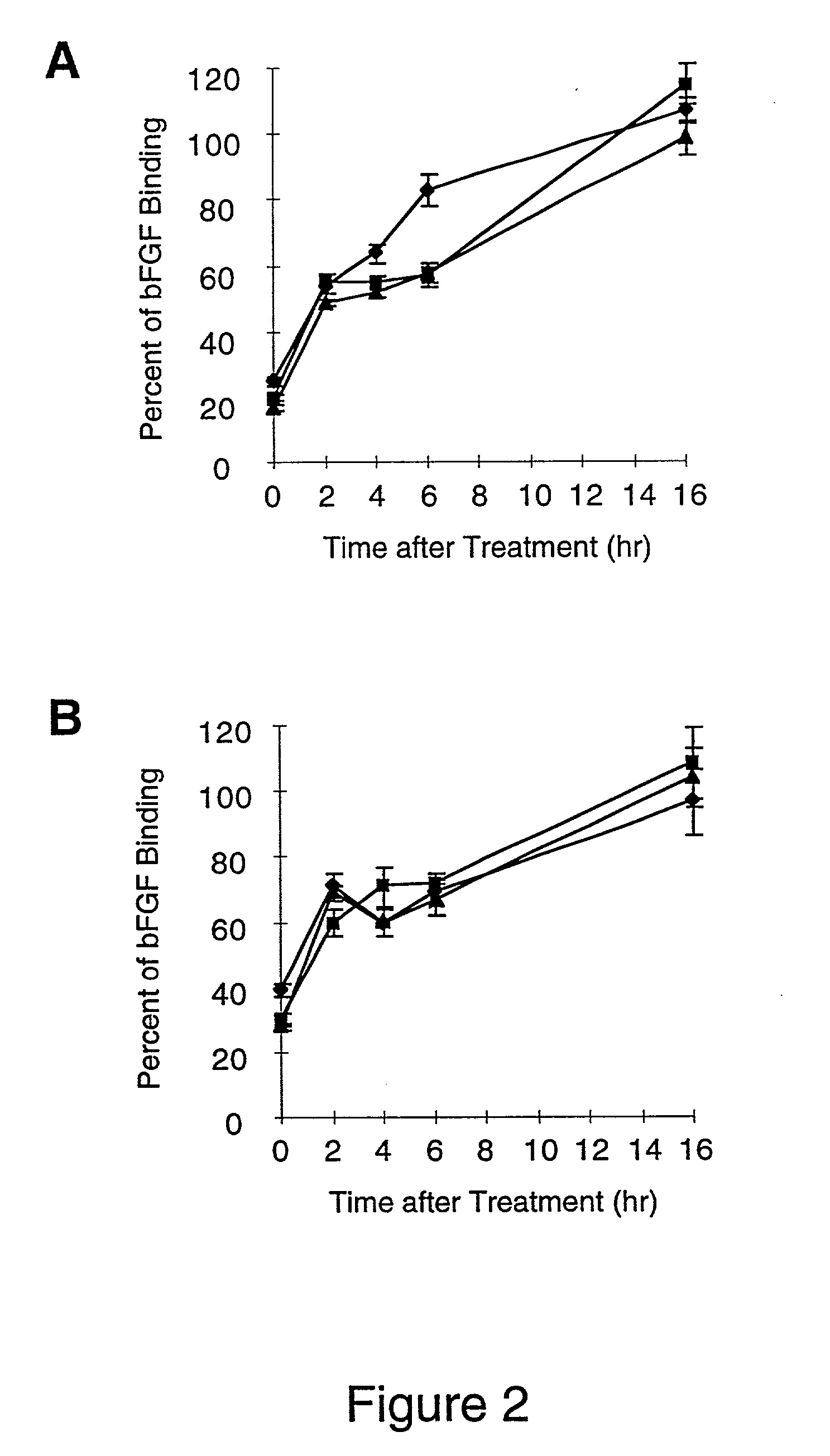
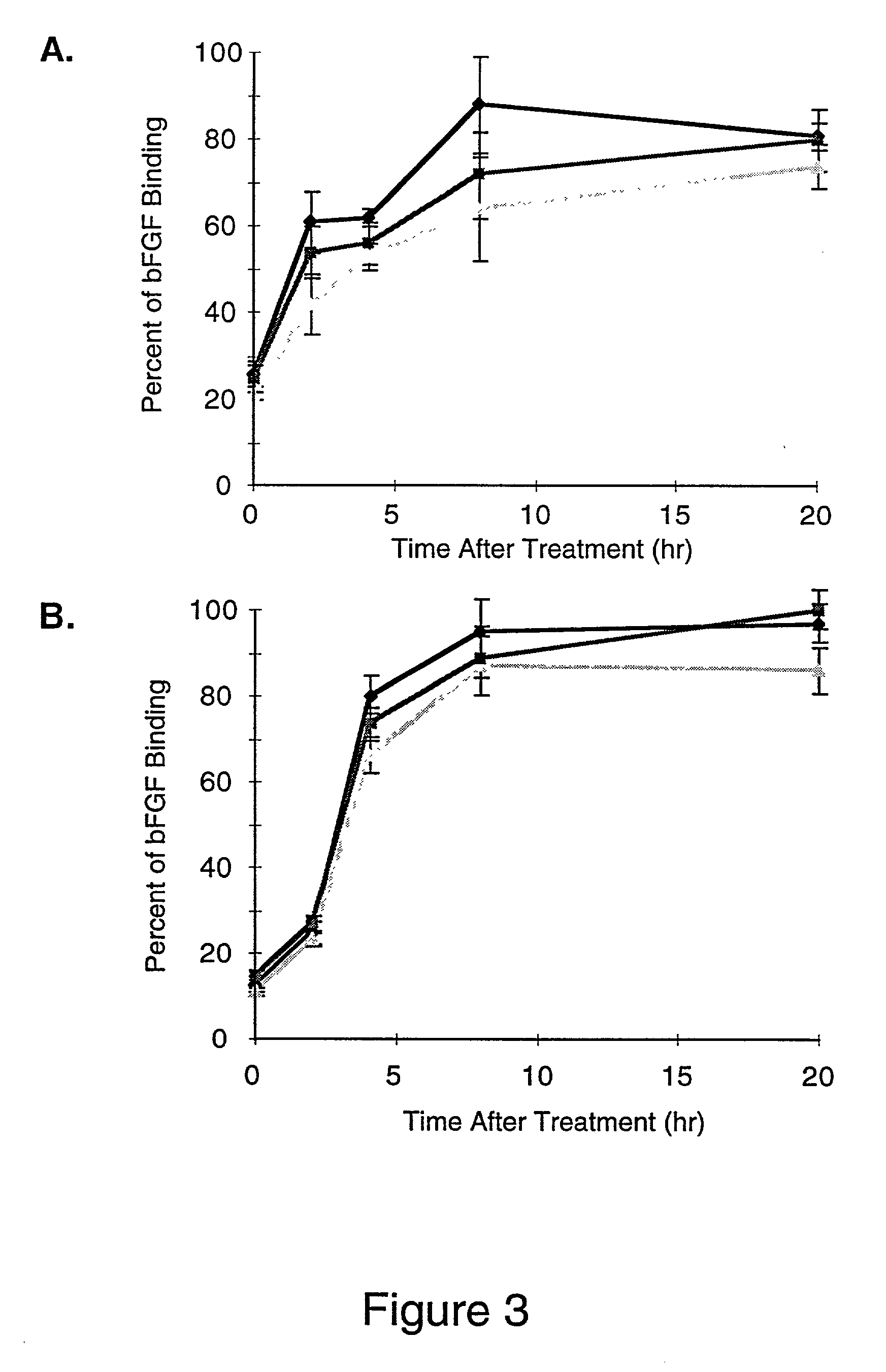
[0071] The growth factor, basic Fibroblast Growth Factor (bFGF), is a well characterized heparin binding protein, which is known to bind to the heparin moieties of proteoglycans on the cell surface and in the extracellular matrix (Maccarana, et al., J. Biol. Chem., 268:23898-23905, 1993). Binding of .sup.125I-labeled bFGF to cell surface and basement membrane proteoglycans was used to access the amount of heparin / heparan sulfate removed from unactivated and IL-1b activated endothelial cell layers and their basement membranes by heparinases I, II or III. Digestion of the cell surface and basement membrane with heparinase will remove both heparin and heparan sulfate moieties, because these moieties are interspersed on the same unbranched carbohydrate chains. .sup.125I-labeled bFGF binding was also used in this experiment to...

example 3

of Activated Endothelial Cell Layers and Basement Membranes with Heparinase to Release Heparin / Heparan Sulfate Bound Chemokine, IL-8

[0075] Removal of the bound chemokine gradient formed by activated endothelium adjacent to inflamed tissue will inhibit the accumulation of neutrophils within this tissue, and will decrease the inflammatory response. The chemokine, IL-8, is produced by endothelium activated by IL-1b and other cytokines and chemoattractants, which are secreted by inflamed tissues. If IL-8 bound to endothelium can be solubilized by treatment with heparinase, then it would be removed from the area of inflammation by blood flow and the localized inflammatory response would be inhibited. The in vitro removal and solubilization of 0.5 to 3 fold more endogenous, immobilized IL-8 (vs. secreted IL-8) from activated endothelium by heparinases I, II or III, or by heparinases I and III demonstrates that the bound chemokine gradient can be destroyed by heparinase treatment.

[0076] On...


PUM

PropertyMeasurementUnit
pHaaaaaaaaaa
pHaaaaaaaaaa
concentrationsaaaaaaaaaa

Abstract

Heparinase enzymes can be used as a medical treatment to reduce localized inflammatory responses. Treatment of activated endothelium with heparinase inhibits leukocyte rolling, adhesion and extravasation. Most of the heparin and heparan sulfate on endothelial cell surfaces and in basement membranes is degraded by exposure to heparinase. In addition, immobilized chemokines, which are attached to heparin/heparan sulfate on activated endothelium are solubilized by heparinase digestion. Heparinase can be infused into the vascular system to inhibit accumulation of leukocytes in inflamed tissue and decrease damage resulting from localized inflammations. Targeting of heparinase to activated endothelium can be accomplished through localized administration and/or use of genetically engineered heparinase containing endothelium ligand-binding domains.

Description

[0001] This application is related to U.S. provisional application Ser. No. 60 / 004,622, filed Sep. 29, 1995, which is expressly incorporated herein by reference.[0002] This invention is in the field of medical treatments and is directed to the use of heparinase enzyme as a treatment or prophylactic for reducing localized inflammatory responses.BACKGROUND OF INVENTION[0003] An inflammatory response is local response to cellular injury that is marked by capillary dilation, leukocytic infiltration, redness, heat, and pain and serves as a mechanism initiating the elimination of noxious agents and of damaged tissue.[0004] A generalized inflammatory response within a tissue occurs by the recruitment of leukocytes to the tissue. Destruction of bacteria, foreign materials and / or damaged cells occurs through phagocytosis and / or extracellular degranulation (secretion of degradative enzymes, antimicrobial proteins and myeloperoxidase, which forms superoxides from secreted H.sub.2O.sub.2). Whil...

Claims


Application Information

Patent Timeline
Patent Type & Authority Applications(United States)
IPC IPC(8): A61K38/00A61K9/14A61K31/00A61K38/46A61K38/47A61K39/395A61K45/00A61K47/48A61P9/10A61P29/00A61P43/00C12N9/24C12N9/88C12N9/96
CPCA61K38/51A61K47/48C07K2319/00C12N9/88Y10S424/81A61K47/50A61P9/10A61P29/00A61P43/00
Inventor BENNETT, D. CLARKCAUCHON, ELIZABETHFINK, DOMINIQUEGROUIX, BRIGETTEHSIA, ARIANEDANAGHER, PAMELAZIMMERMANN, JOSEPH
Owner BIOMARIN PHARMA INC
Why Eureka
Try Eureka