
Locally confined photodynamic treatment for diseased tissue

a tissue and photodynamic treatment technology, applied in the field of local confined photodynamic treatment of diseased tissue, can solve the problems of not teaching the spraying of gels or thermosetting gels onto the treatment site, inefficiency and time-consuming, and the invention is limited to external areas of the skin, so as to achieve efficient uptake of active components.

Inactive Publication Date: 2003-02-06
CERAMOPTEC IND INC
View PDF0 Cites 51 Cited by
  • Summary
  • Abstract
  • Description
  • Claims
  • Application Information

AI Technical Summary

Benefits of technology

[0021] It is yet a further object of the present invention to apply the active composition in a gel form and thereby increase the area which can be treated at one time compared to other locally applied compositions, and to minimize the necessary amount of the photosensitizer while reaching a higher concentration locally than would have been achieved following a systemic administration.
[0024] It is another object of the present invention to provide a device to deliver and evenly distribute the pharmaceutical composition, activate the active component and localize the treatment to a specific desired site for treatment, diagnosis or both treatment and diagnosis.
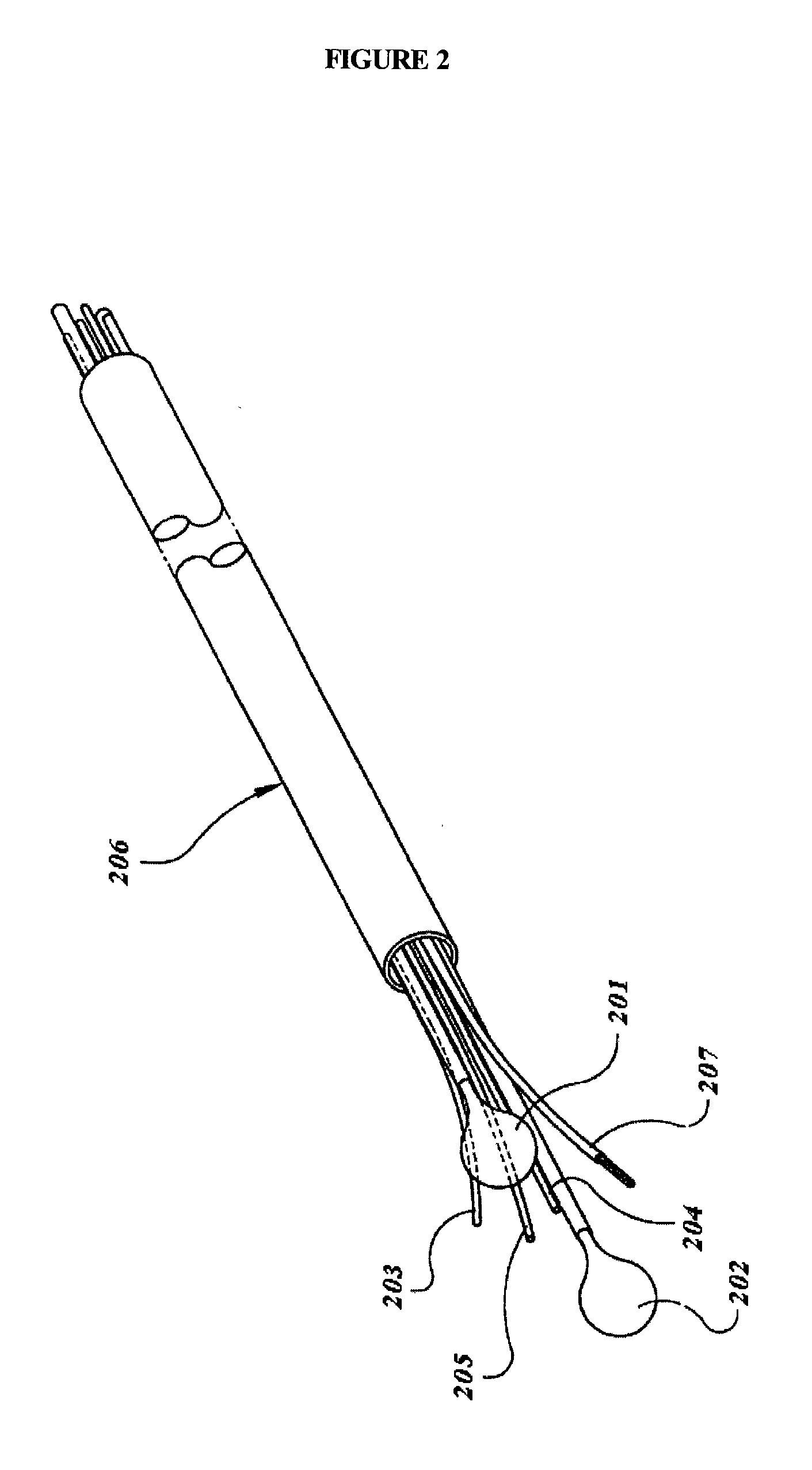
[0025] It is a further object of the present invention to provide a delivery device for the present invention assuring stability of any single components that are mixed directly before the application.

Problems solved by technology

However, neither of these patents demonstrate a formulation that provides a local administration of the active compound to confined tissue areas, particularly in inner organs, with an even distribution of the therapeutic component.
Additionally, it does not teach the spraying of a gel or thermosetting gel onto a treatment site.
This invention is, however, limited to external areas of the skin, and is further limited to applications where stratum corneum is present.
It is therefore unsuitable for lesions of a larger area than the patch area, in that the treatment would have to be administered numerous times to cover the entire lesion, which would be inefficient and time consuming.
Also, the confinement properties of '279 are limited to external use, and would thus not be effective for internal epithelial layers.
With such a narrow beam as compared to the size of the area being treated, the treatment procedure can be time consuming and laborious.
This increased power creates a larger potential for damage to the healthy tissue underneath.
A 2.2 mm spot size can be relatively large for some areas, which would make it difficult to control irradiation along the edge of the diseased area.
This drawback therefore creates a potential for irradiation of surrounding healthy tissue.
This method does not reveal how to treat a larger area more quickly, efficiently and more safely.
However, this method is limited by the lack of depth control in the treating of the tissue.
Freezing depth is difficult to control.

Method used


Image

  • Locally confined photodynamic treatment for diseased tissue
  • Locally confined photodynamic treatment for diseased tissue
  • Locally confined photodynamic treatment for diseased tissue

Examples


example 2

Diagnosis of Skin Diseases

[0053] The pharmaceutical composition was prepared with Lutrol and 5-aminolevulinic acid (ALA) in water. The thermosetting gel was prepared with 17.9% Lutrol in water by stirring under cooling. It was shown that the viscosity of the gel increased with increased concentrations of ALA. Several concentrations in the range of 2 to 40% (w / w) ALA were tested by detecting the fluorescence of protoporphyrin IX on skin. A concentration of 2% ALA only showed very low fluorescence on healthy skin areas compared to diseased skin areas. In general, the fluorescence was very low for this concentration. Therefore, for clinical studies a concentration of ALA of 10% was used, and the duration of incubation was reduced compared to the therapeutic approach (see example 3). The fluid was sprayed onto the areas of the skin to be examined to achieve an even distribution of the active component on a broad area, and the thermosetting gel became highly viscous upon contact with the...

example 3

Treatment of Basal Cell Carcinoma and Actinic Keratosis of the Skin

[0054] The pharmaceutical composition was prepared as described in Example 2. A concentration of 2% ALA only showed very low fluorescence on healthy skin areas compared to diseased skin areas. This is desirable for therapeutic use, however with the low content of ALA one cannot be sure that the active composition penetrates deep enough into the tissue. Therefore and because of the increased viscosity of the gel, higher concentrations of ALA are preferred for therapeutic use. With a concentration of 10% ALA, significantly enhanced fluorescence could be detected which was not significantly increased by concentrations of ALA of 20 or 40%. For the clinical studies, the Lutrol gel with 10% ALA, dissolved directly before use, was applied for 2 hours under light protection. Then the gel was removed and the incubation continued for an additional 2 hours. Lastly, the treatment areas were irradiated at 633 nm with an energy de...

example 4

Treatment of Diseased Tissue at the Uterine Cervix

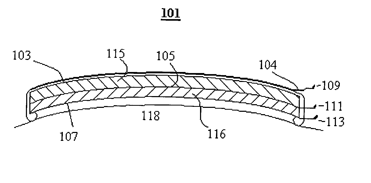
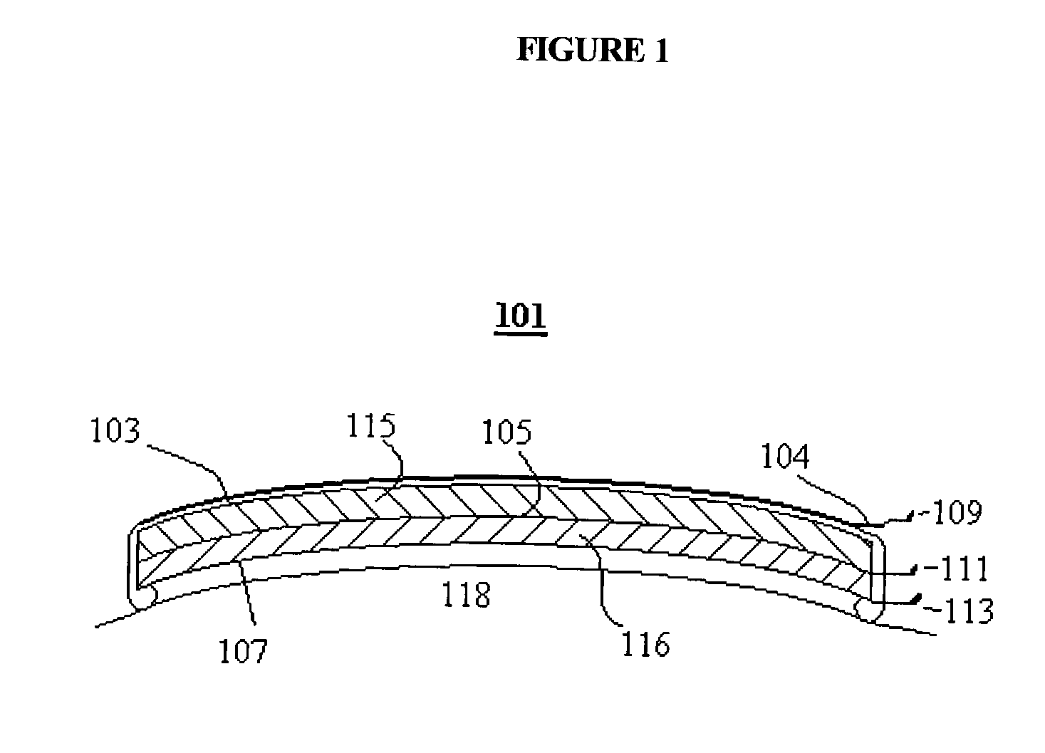
[0055] Treatments of diseased cervical tissue can be performed in a similar fashion as the above examples. ALA is incorporated into gels that are then introduced into the diseased portion of the cervix. In addition to the gel's high viscosity, additional measures are employed to enhance the tendency of the gel to remain localized. In a preferred embodiment, a rubber cup similar to a contraceptive cervical diaphragm is placed in the cervix to further restrict the composition. In another preferred embodiment, a moist dressing especially designed for mucosal surfaces is utilized. After a sufficient uptake period, the additional localization measure and the gel is removed.


PUM

PropertyMeasurementUnit
Viscosityaaaaaaaaaa
Sizeaaaaaaaaaa
Flexibilityaaaaaaaaaa

Abstract

A pharmaceutical composition, method and apparatus for treatment, diagnosis or both treatment and treatment of hyperproliferative malignant and non-malignant diseases of epithelial tissues is disclosed. The invention comprises local application of the pharmaceutical composition to a predetermined area of the tissue characterized by complete and consistent coverage of the tissue, including irregularly shaped tissue. The pharmaceutical composition consists of an active component, such as a photosensitizer or a precursor thereof, and at least one carrier substance including a viscous fluid, a gel, or a fluid that becomes viscous upon contact with the tissue. The gel's viscosity allows it to adhere to the tissue for a sufficient amount of time to transfer the photosensitizer or precursor. In a preferred embodiment the pharmaceutical composition is sprayed onto the surface of the diseased tissue. Optionally, a mechanical device is used to further restrict the composition to a specific areas and can also be used to press the composition onto the tissue. In another embodiment, components of the composition are delivered to and mixed at the treatment site by a suitable delivery device prior to irradiation. The active component is then activated by a suitable wavelength of radiation.

Description

REFERENCE TO RELATED CASE[0001] This application is a continuation of co-pending U.S. patent application Ser. No. 09 / 903,287 filed on Jul. 11, 2001 by Thierry Patrice, Wolfgang Neuberger, Hans-Peter Bode and Ludovic Bourre, inventors, entitled "Treatment for Epithelial Diseases", which in turn was a continuation-in-part of U.S. patent application Ser. No. 09 / 621,802 filed on Jul. 21, 2000 by Thierry Patrice and Wolfgang Neuberger, inventors, entitled "Treatment for Barrett's Syndrome", and incorporated by reference herein.[0002] 1. Field of the Invention[0003] The present invention relates to pharmaceutical compositions, methods and apparatus for diagnosing and treating hyperproliferative pre-malignant, malignant or nonmalignant diseases of the epithelium of an organ or of the skin and tissue lying immediately below the epithelium by PhotoDynamic Therapy (PDT).[0004] 2. Invention Disclosure Statement[0005] Hyperproliferative diseases comprise pre-cancerous or cancerous states or vir...

Claims


Application Information

Patent Timeline
IPC IPC(8): A61B17/22A61B18/00A61B18/22A61N5/06
CPCA61B18/22A61B2017/22054A61B2018/00982A61B2018/2261A61K9/0014A61K47/32A61K47/34A61N5/0601A61N5/062A61N2005/0602A61N5/0603A61B2018/00559
Inventor NEUBERGER, WOLFGANGSPANIOL, STEFANBODE, HANS-PETERPATRICE, THIERRYBOURRE, LUDOVIC
Owner CERAMOPTEC IND INC
Learn More