[0005] The object of the present invention is to provide a reagent and a method for an immunological analysis of elastase 1, which can be analyzed conveniently without a special equipment, quickly so that an emergency examination can be carried out, and quantitatively in a wide range from a low concentration range to a
high concentration range.
[0022] As smaller latex particles, for example, latex particles having a particle size of preferably 0.05 to 0.3 .mu.m, more preferably 0.05 to 0.25 .mu.m may be used. As larger latex particles, for example, latex particles having a particle size of preferably 0.2 to 0.5 .mu.m, more preferably 0.25 to 0.5 .mu.m may be used. A combination of latex particles having the above particle sizes is used in the present invention, and thus a detectable limit at the side of a low concentration can be improved and a good reproducibility can be obtained. At the side of a
high concentration, misunderstanding caused by a decrease of an
agglutination property (i.e., prozone phenomenon in which an
agglutination property is decreased due to an excess
high concentration, agglutination is seemingly decreased, and a measured value lower than an actual value is obtained) can be avoided, even if a concentration of elastase 1 is high.
[0040] The measurement of elastase 1 has advantages in comparison with conventional markers for a
pancreatic disease. For example, when suffering from a
stomach ache but visiting a hospital after several days therefrom (i.e., not an emergency case), conventional markers such as
amylase or
lipase are often normalized. In contrast, a biological half-life of elastase 1 in blood is so long that, if elastase 1 shows an abnormal value when visiting, the abnormal value may be an evidence (a late marker) which shows a disorder of
pancreas (or
pancreatic duct) at least several days ago, and thus it becomes necessary to carry out a clinical examination, diagnosis, and treatment, including a possibility of a
pancreatic cancer. In other words, a period from the alleviation of an acute symptom to normalization with respect to elastase 1 is longer (for example, a week to a month) than that (several days) of
amylase or
lipase. From this point, the present invention in which elastase 1 can be measured quickly, conveniently, and with sensitivity has an excellent availability.
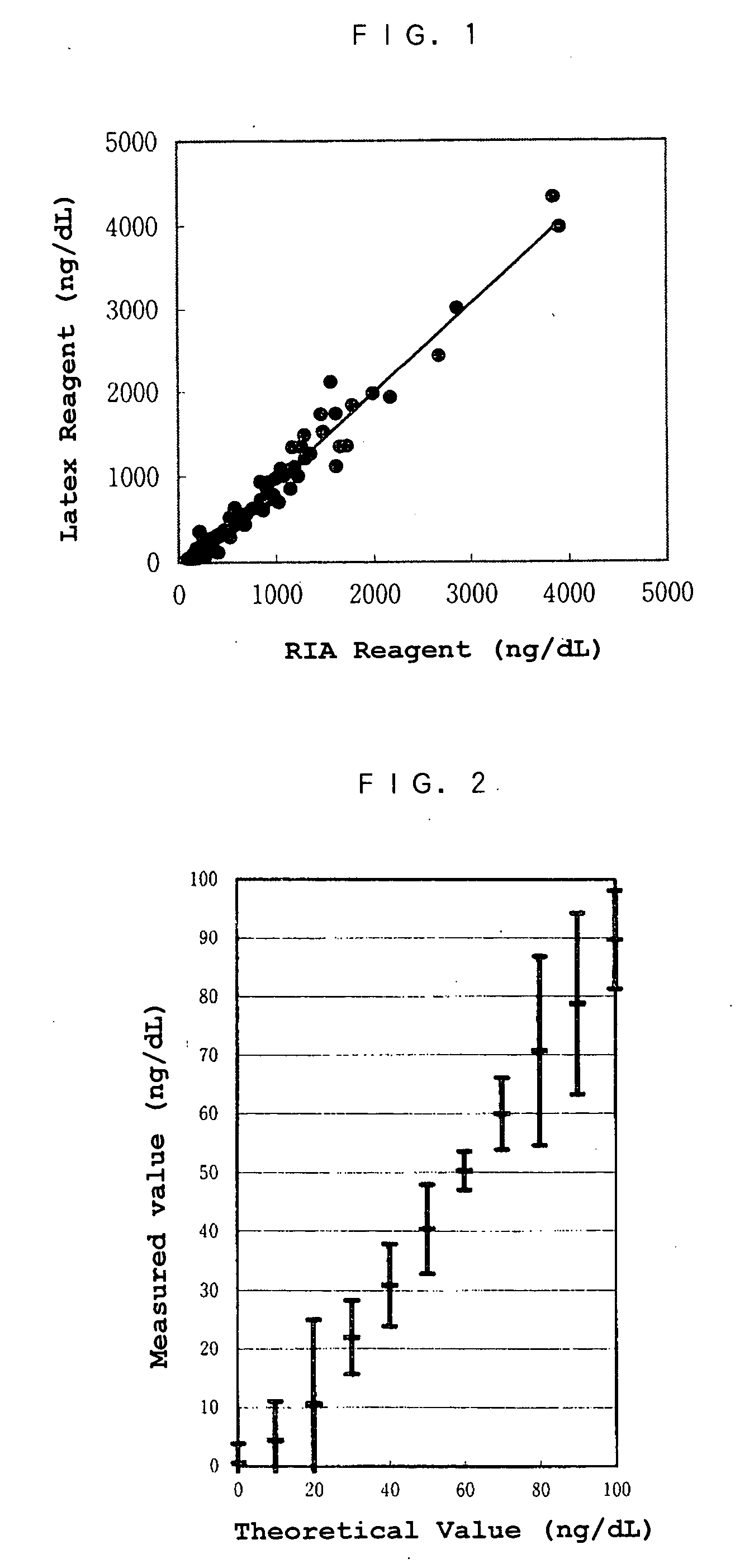
[0041] For example, an accurate measurement at or near the above concentration (400 ng / dL) is necessary to judge the existence of
acute pancreatitis. Such a concentration cannot be accurately measured by an EIA method, and thus only an RIA method can be selected among conventional methods. However, a measurement accuracy (reproducibility) is not sufficient at the above concentration range even in the RIA method. The above problems can be solved by the present invention. Because of the use of a latex method, elastase 1 can be accurately measured in a wide range, and the present invention has a remarkable effect in that a pancreatic
disease can be accurately detected.
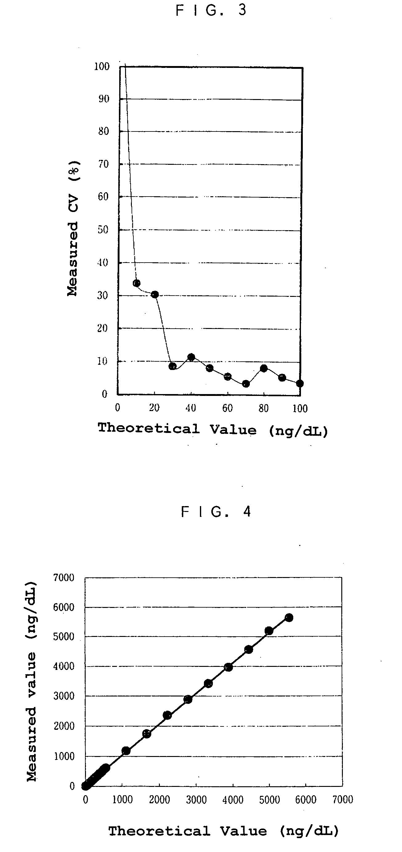
[0042] According to the present invention, in which a low value of elastase can be accurately measured, chronic
pancreatitis can be accurately monitored, and thus various pancreatic diseases can be widely judged. In the past, the monitoring of chronic
pancreatitis has depended on amylase, which is not exactly specific to pancreas. According to the present invention, elastase 1 from low values to high values, which cannot be measured by a conventional method, can be measured conveniently and quickly, without an
additional procedure such as a
dilution of a sample or increase of an amount applied. The above effects have excellent advantages in the field of clinical examination.
[0043] As described above, the reagent for measuring elastase 1 by a latex agglutination
reaction system according to the present invention is novel, and a pancreatic
disease can be accurately detected by accurately measuring elastase 1 using the latex agglutination reagent.

