High-density cell microarrays for parallel functional determinations

a high-density cell and functional determination technology, applied in the field of high-density cell microarrays, can solve the problems of difficult to achieve using conventional techniques, difficult to rapidly uncover gene regulatory circuits and functional manifestations at the cellular level, and difficult to study phenotypic manifestations

Inactive Publication Date: 2005-01-20
MEMORIAL SLOAN KETTERING CANCER CENT
View PDF2 Cites 6 Cited by
  • Summary
  • Abstract
  • Description
  • Claims
  • Application Information

AI Technical Summary

Benefits of technology

[0004] We have developed methods for constructing high-density cell microarrays and have demonstrated that these microarrays allow for phenotypic determinations of gene activities on a large scale. Specifically, we have found that the generation of nanocraters ranging in size from about 100 pico-liters to 1.5 nano-liters on permeable membranes allows for the creation of high-density cell microarrays. Cells inoculated into the nanocraters form individual colonies that remain confined to the nanocraters and can, therefore, be arrayed very closely together (i.e. at high density) without cross-contamination.

Problems solved by technology

However, there are still several challenges to studying phenotypic manifestations of gene activities on a genomic scale.
In particular, large-scale phenotypic analyses require that cells be grown in parallel and in miniaturized format without cross-contamination, which can be difficult to accomplish using conventional techniques.
Thus, although whole-genome sequencing projects have generated a wealth of gene sequences from a variety of organisms, developing methods for rapidly uncovering gene regulatory circuits and their functional manifestations at the cellular level remains a major challenge.

Method used


Image

  • High-density cell microarrays for parallel functional determinations
  • High-density cell microarrays for parallel functional determinations
  • High-density cell microarrays for parallel functional determinations

Examples


example 1

Preparation of E. coli Cell Microarrays

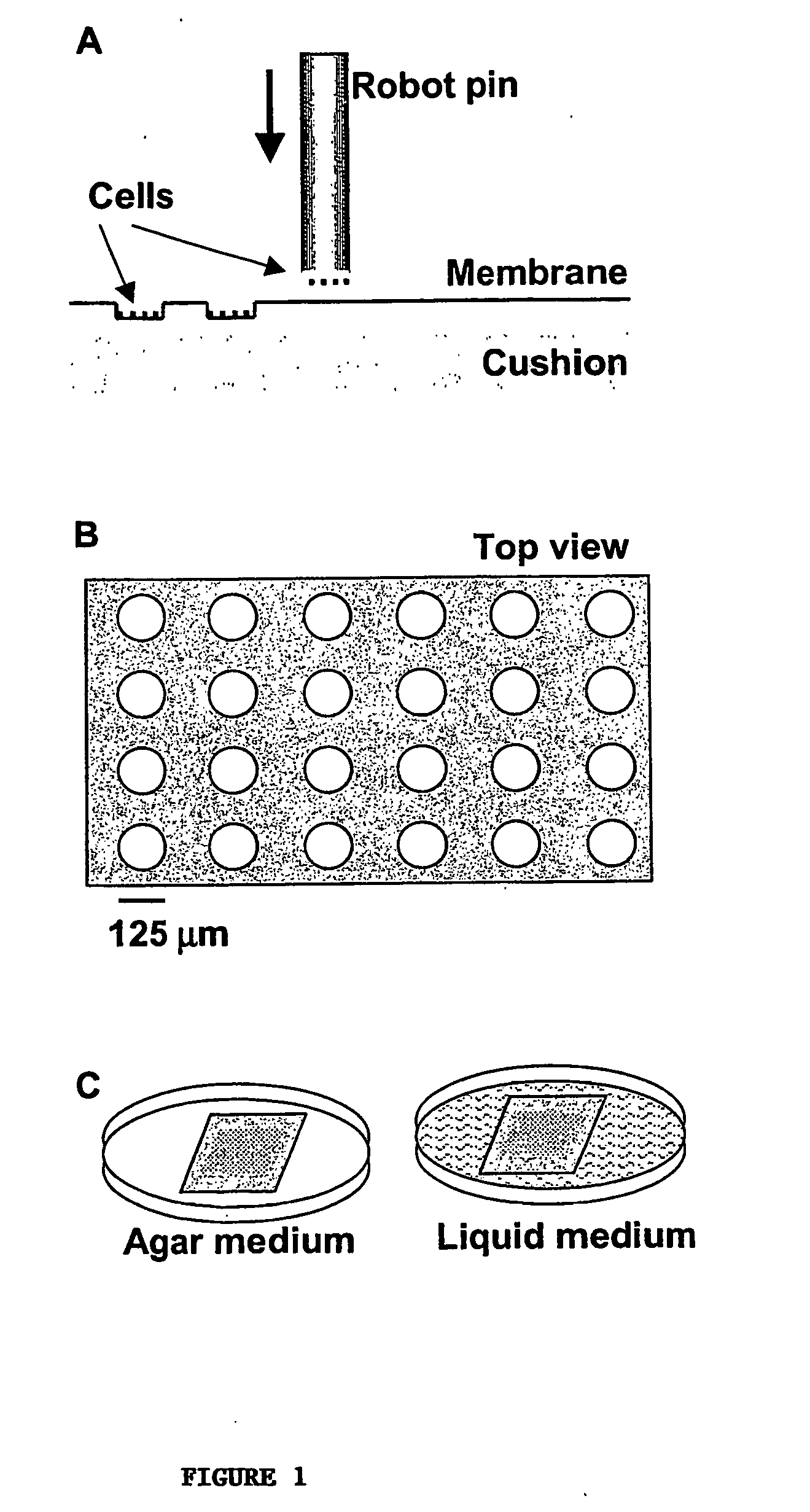
[0041] To illustrate the coupled fabrication process, we first made cell microarrays of E. coli that express β-galactosidase. A cellulose ester membrane (Spectrum) was rinsed with and stored in deionized water at 4° C. until use. The molecular weight cut off for the membrane was 3,500 Daltons and the thickness was estimated to be 10 μm. The membrane was placed on the surface of chromatography paper (Fisher) that had been soaked in a warm 0.5% agarose solution and placed on a standard microscope slide. This cushion helped to immobilize and moisturize the membrane during the arraying process. Any bubbles or excessive agarose between the cushion and the membrane was removed by gently rubbing the membrane with a clean and smooth rod. The membrane assembly was then placed in the slide holder of a robotic arrayer (GMS417, Affymetrix).

[0042] An overnight bacterial culture was dispensed into a 96-well plate (Corning) and arrayed by the robot onto the...

example 2

Preparation of S. cerevisiae Microarrays

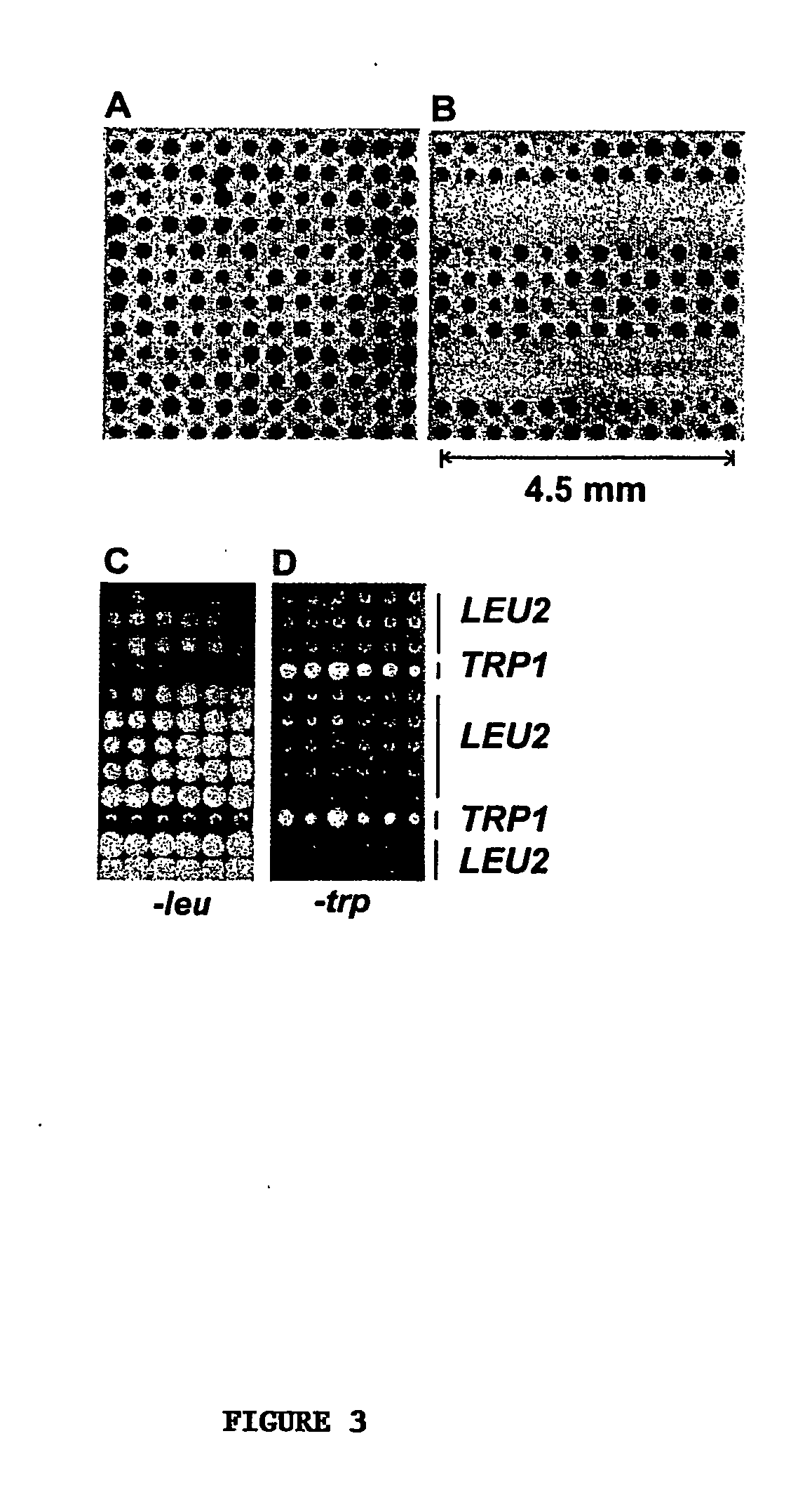
[0045] Using the coupled fabrication approach described in Example 1, we next developed yeast (S. cerevisiae) cell microarrays. Two-day yeast cultures (1.2 mL) in 96-tube format (VWR) were centrifuged at 3,000 rpm for 5 min. The clear supernatant was quickly decanted without perturbing the cell pellets. About 20 μL of concentrated yeast was transferred to a 96-well plate (alternatively, the yeast cells can be resuspended in YPD+15% glycerol before arraying). Yeast cultures were dispensed into a 96-well plate and arrayed using a robotic arrayer as described above.

[0046] Shown in FIGS. 3C and 3D are yeast cell microarrays of an auxotrophic strain carrying either LEU2 or TRP1 plasmids. The cell array membranes were placed onto the surface of synthetic media lacking either leucine or tryptophan and incubated at 30° C. for 12-24 hours. Yeast cells with a LEU2 plasmid grew in the medium lacking leucine while those with a TRP1 plasmid did not. Conv...

example 3

S. cerevisiae Cell Microarrays for Identifying Drug Targets

[0047] To further illustrate the utility of cell microarrays for assaying drug effects on individual gene deletions, we used a series of diploid strains carrying homozygous gene deletions of fkb1 and 93 other genes chosen at random. (Winzeler, E. A. et al. Functional characterization of the S. cerevisiae genome by gene deletion and parallel analysis. Science 285, 901-906. (1999)). FKB1 encodes FKBP12 that binds FK506 and rapamycin, two natural products used as anti-fungal and immunosuppressant drugs. The FKBP-drug complex inhibits progression through the G1 phase of the cell cycle in yeast and mammalian cells. Deletion of FKB1 has been shown to render yeast resistant to rapamycin. (Heitman, J., Movva, N. R. & Hall, M. N. Targets for cell cycle arrest by the immunosuppressant rapamycin in yeast. Science 253, 905-909. (1991); Schreiber, S. L. & Crabtree, G. R. Immunophilins, ligands, and the control of signal transduction. Ha...


PUM

PropertyMeasurementUnit
distanceaaaaaaaaaa
distanceaaaaaaaaaa
distanceaaaaaaaaaa

Abstract

Disclosed are methods for generating high-density cell microarrays. The methods generally involve forming nanocraters on a permeable membrane surface and inoculating the nanocraters with cells, proteins, or other molecules. The high-density microarrazs of the invention are useful for large-scale, high throughput phenotypic determinations of gene activities.

Description

FIELD OF THE INVENTION [0001] The invention relates to high-density cell microarrays and methods for their preparation and use. BACKGROUND OF THE INVENTION [0002] The availability of full genome sequences has generated great interest in studying gene functions on a genome-wide scale. Technologies are being developed that allow for the global analysis of important macromolecules that convey the information flow from DNA to RNA to proteins in cells. For example, DNA microarrays have allowed gene expression profiling for specific cellular states, and global two-hybrid analyses have provided a glimpse of intracellular signal wiring systems in yeast. Biochemical genomics will eventually enable the genome-wide analyses of protein activities, and genomic surveys of the targets of DNA binding proteins are certain to have an impact on our understanding of regulatory circuits. Systematic gene deletion projects have also begun to provide insights into gene function on a large scale. [0003] Sin...

Claims


Application Information

Patent Timeline
Patent Type & Authority Applications(United States)
IPC IPC(8): C40B60/14G01N15/14G01N35/00
CPCB01J2219/00317G01N2035/00158B01J2219/00387B01J2219/00527B01J2219/00603B01J2219/00641B01J2219/00691B01J2219/00743C40B60/14G01N15/1456G01N15/1463G01N15/1475G01N35/00029G01N2015/1477G01N2015/1497B01J2219/00382
Inventor XU, WILSON C
Owner MEMORIAL SLOAN KETTERING CANCER CENT
Why Eureka
Try Eureka