

Methods and compositions for the assessment of polymer assembly
a polymer and assembly technology, applied in the field of methods for the assessment of polymer assembly, can solve the problems of serious clinical consequences, pathologic conditions, and the inability of the technology for investigating the assembly of extracellular matrix to provide a means for real-time investigation of cell-mediated extracellular formation of molecules
- Summary
- Abstract
- Description
- Claims
- Application Information
AI Technical Summary
Benefits of technology
Problems solved by technology
Method used
Image

Examples
example 1
Vascular Smooth Muscle Cell Isolation and Culture
[0081] Mouse aortic smooth muscle cells (SMC) were isolated from explants of aortas harvested from mice in the 129 SvEv genetic background. Briefly, the aortas were excised, the adventitia was removed, the aorta cut into approximately 1-mm rings, and the rings plated on scored areas of a Petri dish. After 2 weeks, the aortic rings were removed and the cells were grown for an additional 2 weeks before passaging. The primary culture and the first passage of SMC were grown in DMEM supplemented with 20% FBS, 1% L-glutamine and penicillin-streptomycin, followed by DMEM with 10% FBS for subsequent cultures.
[0082] An explant method also was used to isolate SMC from human saphenous vein. Human saphenous vein segments, obtained as excess after coronary bypass surgery, were cut longitudinally, unfolded, and adventitia and intima were removed using blunt dissection with forceps. The media was minced and plated onto Petri dishes, as described a...
example 2
Protein Labeling
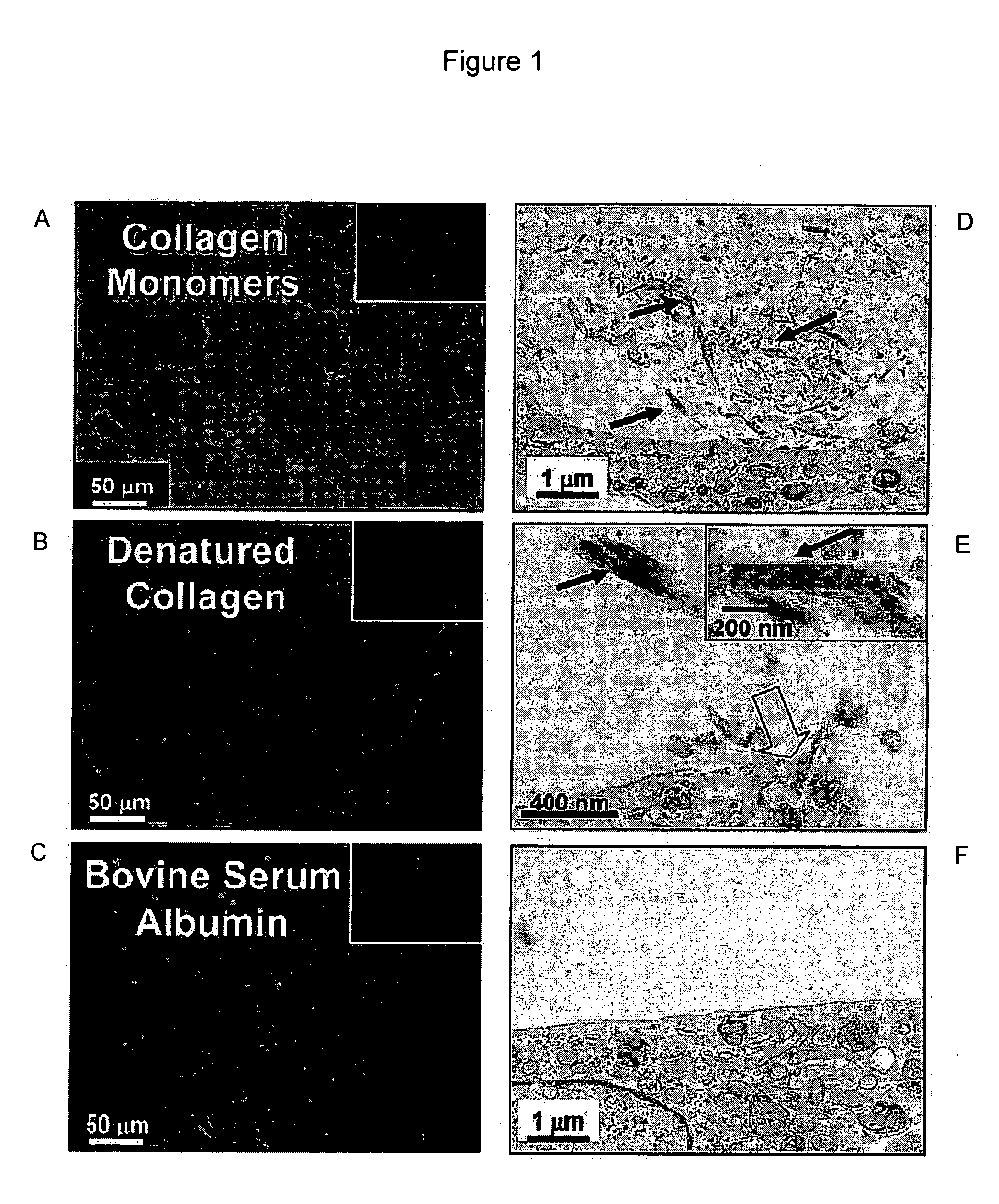
[0083] To verify the ability of the assay to discriminate cell-mediated protein assembly, collagen monomers were compared with BSA and denatured collagen. A bovine serum albumin (BSA) solution (4 mg / mL in deionized water) and acid solubilized rat-tail or bovine-skin collagen were dialyzed separately against borate-buffered saline (100 mM of sodium borate, 1 M of sodium chloride, pH 9.3) using dialysis tubing (8000 MWCO, Spectrum Medical Industries), for 24 h at 4° C.
[0084] At this pH, necessary for the fluorescein labeling, the concentrated collagen solution gelled. The dialyzed protein solutions were transferred to reaction vessels to which 7.9 L of FLUOS [Roche, 20 mg / mL fluorescein isothiocyanate (FITC) in DMSO] per mg of protein were added (an approximately 56:1 molar ratio of FLUOS to protein). The labeling mixture was allowed to react for 3 h at room temperature with gentle stirring and protected from light.
[0085] After labeling, the FLUOS-protein mixture wa...
example 3
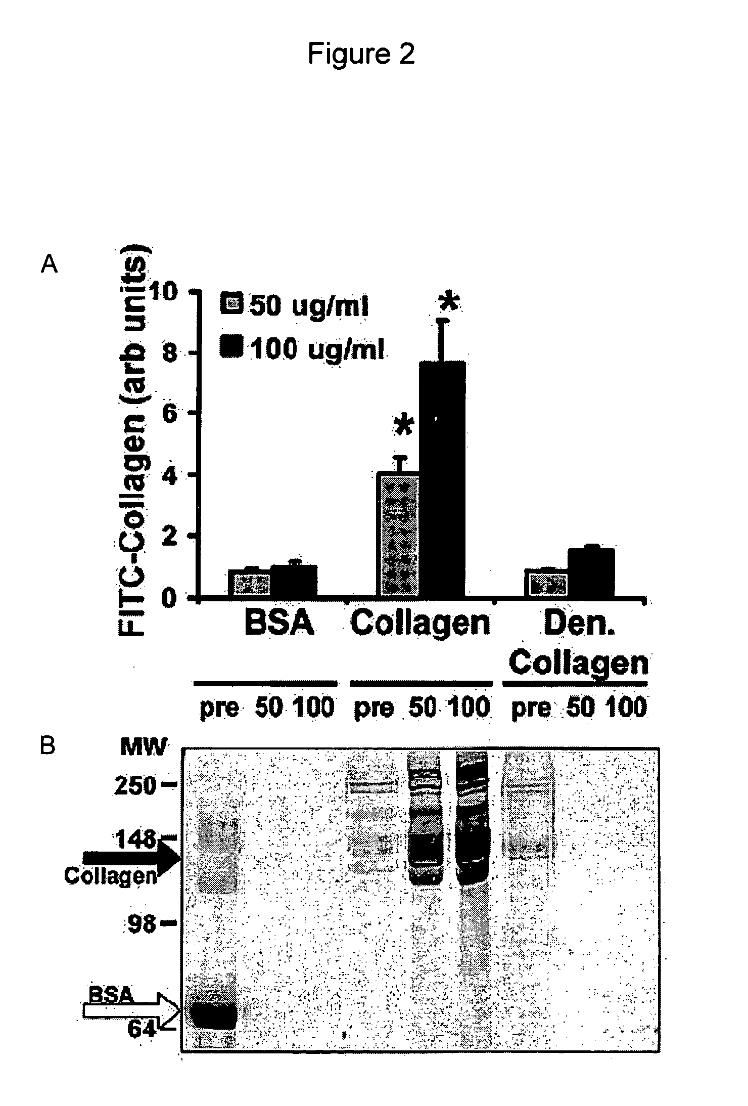
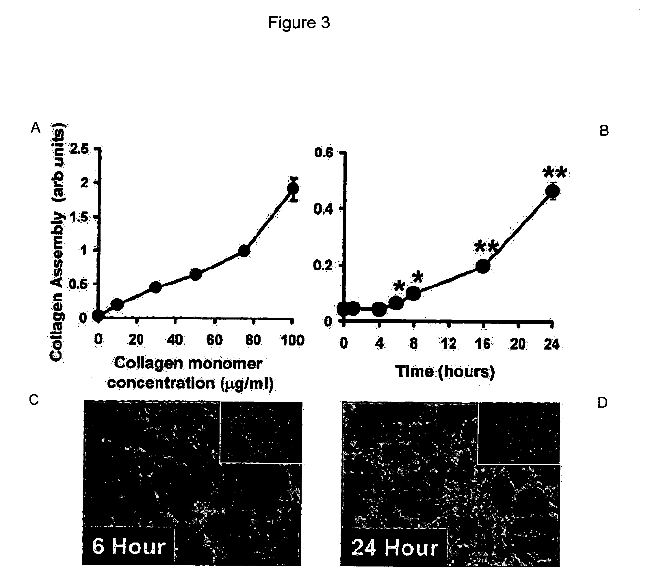
[0088] In one embodiment, the assay consists of adding a solution of FITC-labeled protein to cultured cells in the presence or absence of various treatments, then returning the cells to the incubator (37° C., 5% CO2) for various periods of time. Supramolecular protein assembly can be followed in time as a gain of fluorescence using a fluorescence plate reader, or by confocal microscopy with live cells or after fixation of cells. Specifically, for the development of this assay, SMC were used that had been plated into black clear-bottomed 96-well tissue culture-treated plates (Fisher) at 10,000 cells per well in 100 L of DMEM+10% FBS 2 days prior to performing the assay.
[0089] After 24 h, the culture medium was changed to DMEM without FBS to promote cell quiescence, then SMC were returned to the incubator (37° C., 5% CO2) for an additional 24 h. Before the start of the assay, the culture medium was aspirated from each well and replaced with 100 L of FITC-labele...
PUM
| Property | Measurement | Unit |
|---|---|---|
| Time | aaaaa | aaaaa |
Abstract
Description
Claims
Application Information