

Liposome-containing radiographic contrast medium and preparation method thereof
- Summary
- Abstract
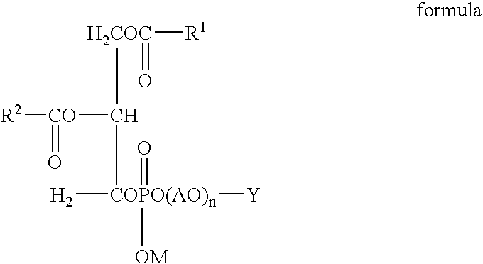
- Description
- Claims
- Application Information
AI Technical Summary
Benefits of technology
Problems solved by technology
Method used
Image

Examples
example 1
[0074] Dipalmitoylphosphatidylcholine (DPPC) and carbon dioxide were added together with ethanol into a stainless steel autoclave and stirred with maintaining the autoclave at 60° C. and 300 kg / cm2 to dissolve the DPPC in supercritical carbon dioxide. Iomeprol solution was continuously added using a metering pump, while stirring the supercritical carbon dioxide solution. The iomeprol solution was prepared in such a manner that 816.5 mg of iomeprol was dissolved in water for injection with heating, ascorbic acid was added in an amount of 20 mM and 1 mg of tromethamol was further added; the pH was adjusted to a physiological pH and finally, water for injection was added to make up 1.0 ml. Thereafter, the autoclave was evacuated to discharge carbon dioxide and a dispersion of a liposome containing iomeprol was obtained. The obtained dispersion was put into a glass vial and subjected to autoclave sterilization at 121° C. for 20 min to obtain a contrast medium.
[0075] The vesicular parti...
example 2
[0076] The contrast medium obtained in Example 1 was diluted with an isotonic glucose solution to a concentration of 50 mg iodine / ml. When this solution was given to a rat by an intravenous injection, concentration to the liver was observed in radiography. It was noted that the imaging level (or image contrast) in the liver decreased in parallel to imaging levels of all other organs over an elapse of time, and almost all of the iomeprol was discharged into urine.
example 3
[0077] The contrast medium obtained in Example 1 was diluted with an isotonic glucose solution to a concentration of 50 mg iodine / ml. This solution was injected into a vein of a rat having a large number of transferred liver cancer cells. The transferred tumor region exhibited a high imaging level (high image contrast), in which tumors of 5 mm in diameter were observed. Lowering of the image contrast in the tumor region was delayed over an elapse of time, compared to imaging levels of other organs.
PUM
| Property | Measurement | Unit |
|---|---|---|
| Temperature | aaaaa | aaaaa |
| Length | aaaaa | aaaaa |
| Length | aaaaa | aaaaa |
Abstract
Description
Claims
Application Information