
Poly (L-glutamic acid) paramagnetic material complex and use as a biodegradable MRI contrast agent

a paramagnetic material and complex technology, applied in the field of polymer complexes, can solve the problems of inability to achieve the effect of contrast agents without the use of contrast agents, inability to use angiography, and increased risk of patients, and existing formulations of chelated gd are not suitable for use as contrast agents for blood pool imaging

Inactive Publication Date: 2005-07-14
BOARD OF RGT THE UNIV OF TEXAS SYST
View PDF31 Cites 32 Cited by
  • Summary
  • Abstract
  • Description
  • Claims
  • Application Information

AI Technical Summary

Benefits of technology

[0031] The number average molecular weight of the PG polymer complex may be between approximately 10,000 and 500,000. In more specific embodiments, it may be between approximately 50,000 and 300,000.

Problems solved by technology

In many cases this cannot be achieved without the use of contrast agents because non-enhanced imaging techniques are useful only when relatively large tissue areas are involved in a disease process.
Existing techniques to image blood vessels include angiography which is an invasive technique that may be painful, expensive and of increased risk to a patient than a noninvasive imaging technique.
Gd and Mn provide sufficient changes in relaxivity for MRI, however, because of toxicity and poor solubility of free paramagnetic heavy metal cations at physiologic pH, chelated complexes are used to decrease toxicity.
Existing formulations of chelated Gd are not suitable for use as contrast agents for blood pool imaging due to their partial leakage through the vessel walls (extravasation into the extravascular space), their rapid elimination through the kidneys, or their general toxicity, among other problems.
Although polymers have previously been combined with paramagnetic materials in MRI contrast agents, many polymers suffer from a variety of problems when used in this manner.
Dextran, dendrimers and polyamides have also been used as polymeric blood-pool contrast agents but these materials are not biodegradable, leaving ample room for improvement.
However, early spread of tumors to lymph nodes may evade detection because traditional clinical evaluation of lymph node metastases are not very sensitive, and therefore patients often undergo unnecessary removal of entire regional nodal basins for precautionary pathological assessment.
However, LSNB remains an invasive procedure, the results of which may not be known for up to two weeks.

Method used


Image

  • Poly (L-glutamic acid) paramagnetic material complex and use as a biodegradable MRI contrast agent
  • Poly (L-glutamic acid) paramagnetic material complex and use as a biodegradable MRI contrast agent
  • Poly (L-glutamic acid) paramagnetic material complex and use as a biodegradable MRI contrast agent

Examples


example 1

Materials and Analytical Methods

[0091] In these examples, materials were obtained from the following sources: PG sodium salt; N-tert-butoxycarbonyl-1,6-diaminohexane hydrochloride (t-Boc-Hex-NH2); 1,3-diisopropylcarbodiimide; pyridine; 4-dimethylaminopyridine; trifluoroacetic acid (TFA); diethylenetriaminepentaacedic acid (DTPA) dianhydride; gadolinium (III) chloride hexahydrate; 2,4,6-trinitrobenzenesulfonate (TBNS); 4-(2-pyridylazo)resorcinol (PAR); PBS (0.01 M phosphate buffered saline containing 138 mM NaCl and 2.7 mM KCl, pH 7.4) Cathepsin B and all other reagents not otherwise indicated in this example and solvents were purchased from Sigma Aldrich (St. Louis, Mo.).

[0092] Succinimidyl ester of methoxy poly(ethylene glycol)propionic acid (mPEG-NHS) was purchased from Shearwater (Huntsville, Ala.).

[0093]111InCl3 was obtained from Perkin-Elmer Life Sciences (Boston, Mass.).

[0094] p-Isothiocyanatobenzyl-diethylenetriaminepentaacetic acid (p-SCN-Bz-DTPA′3HCl) and p-aminobenzyl-...

example 2

Synthesis of 6-aminohexyl Poly(L-glutamine)

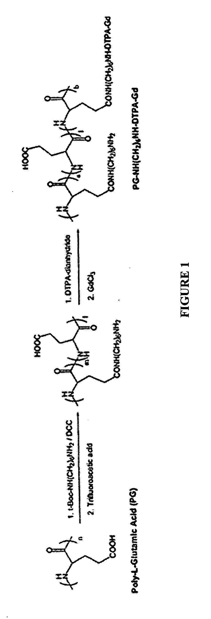
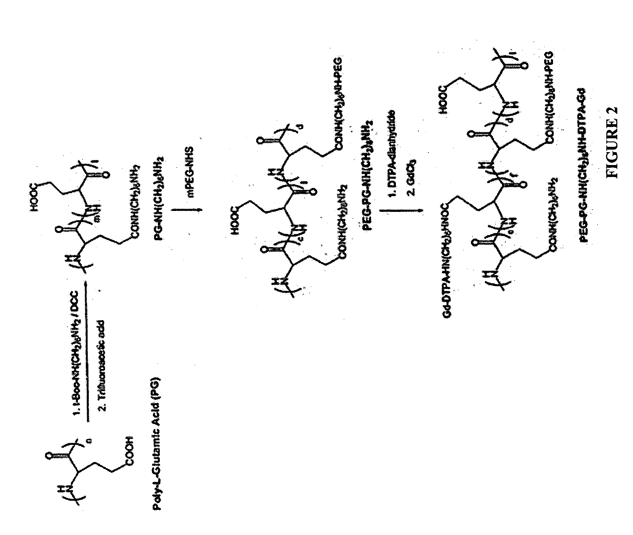
[0100] As part of a method of the present invention, PG functionalized with one or more 6-aminohexyl groups was prepared. The resulting molecule was of the general formula PG[-NH(CH2)6NH2]n, where n is greater than or equal to one. As shown in FIG. 1, the 6-aminohexyl group is attached to a side chain of a glutamic acid residue via the carboxyl group.

[0101] To prepare the 6-aminohexyl functionalized PG first an aqueous solution of sodium salt of PG with a number average molecular weight (Mn) of 17,000 and a degree of polymerization of 116 was converted to its acid form by acidifying with 1 N HCl to pH 3-4. The polymer precipitate was collected by centrifugation, washed with deionized water and lyophilized. 1.3 g of the lyophilized polymer was next added to 20 ml of anhydrous dimethylformamide to prepare a 10 mmol [COOH] solution. To this solution was added 2.0 g (8 mmol) of t-Boc-Hex-HN2 hydrochloride, 1.0 g (8 mmol) 1,3-diisopropylcarbod...

example 3

Synthesis of PG-Hex-DTPA-Gd

[0102] Also as shown in FIG. 1, the DTPA of PG-Hex-DTPA-Gd is conjugated to the 6-aminohexyl group of the PG side chains. To prepare a PG-Hex-DTPA-Gd complex, an approximately 1.1 mmol [NH2] solution of 6-aminohexyl PG prepared as described above was prepared by dissolving 500 mg of the compound in 15 ml of 0.1 M NaHCO3. To this solution, 1.43 g of DTPA-dianhydride (4 mmol) was added in portions over a period of 30 minutes. The pH of the reaction solution was adjusted to 8 by adding aliquots of 0.1 N NaOH solution over the 30 min period. After stirring at room temperature for two hours, the reaction mixture was dialyzed against PBS and deionized water at MWCO 10,000. The resulting solution was concentrated to 5 ml on a centrifugal filter at MWCO 10,000 and stored at 4° C. for future use. The amount of DTPA attached to the PG was determined by quantifying unreacted amino groups with a TNBS assay. Approximately 60% of the amino groups available at the start...


PUM

PropertyMeasurementUnit
flow rateaaaaaaaaaa
diameteraaaaaaaaaa
diameteraaaaaaaaaa

Abstract

The present invention includes PG polymer complexes with paramagnetic materials, such as Gd, Mn and iron oxide. The complexes may also include chelating agents which may be covalently attached to the PG polymer backbone through linkers. PEG may also be attached to the PG polymer backbone. The complexes may include targeting molecules. The complexes are useful as MRI contrast agents, particularly as blood pool agents.

Description

PRIORITY CLAIM [0001] The present application claims priority under 35 U.S.C. §119(e) to U.S. Provisional Patent Application 60 / 532,555, filed Dec. 24, 2003, titled “Poly (L-Glutamic Acid) Paramagnetic Material Complex and Use as Biodegradable MRI Contrast Agent”.STATEMENT OF GOVERNMENT INTEREST [0002] Portions of the present invention were developed using funding provided by the National Institutes of Health, Contract Number U54 CA90810. The U.S. Government has certain rights in the invention.FIELD OF THE INVENTION [0003] The present invention relates in general to paramagnetic material and polymer complexes. Additionally, the invention relates to magnetic resonance imaging (MRI) and in specific to use of paramagnetic material and polymer complexes as MRI contrast agents for blood pool imaging that has more prolonged contrast enhancement of the vasculatures-arteries, venin, inflammation, infection, tumor, etc. than Gd-DTPA. BACKGROUND OF THE INVENTION [0004] Medical diagnostic imag...

Claims


Application Information

Patent Timeline
Patent Type & Authority Applications(United States)
IPC IPC(8): A61K49/00A61K49/08A61K49/14A61K51/08
CPCA61K49/085A61K49/146A61K49/126
Inventor LI, CHUNWEN, XIAOXIAJACKSON, EDWARD F.WALLACE, SIDNEY
Owner BOARD OF RGT THE UNIV OF TEXAS SYST
Learn More