Dual-Modality PET/MRI Contrast Agents

a contrast agent and dual-modal technology, applied in the direction of radioactive preparation forms, pharmaceutical delivery mechanisms, therapy, etc., can solve the problems of false diagnosis, complex attachment process of additional radioactive isotopes, and limitations of the techniques described above, and achieve excellent magnetic properties, remarkable imaging ability, and effective and stably attached

Inactive Publication Date: 2011-05-26
IND ACADEMIC CORP FOUND YONSEI UNIV +1
View PDF3 Cites 14 Cited by
  • Summary
  • Abstract
  • Description
  • Claims
  • Application Information

AI Technical Summary

Benefits of technology

[0017]It is an object of this invention to provide an effective dual-modality PET (positron emission tomography)/MRI (magnetic resonance imaging) contrast agent. Therefore, the present invention utilizes a magnetic signal generating core with excellent magneti

Problems solved by technology

However, current single imaging modality methods tend to be not adequate, resulting in false diagnosis.
However, the techniques described above have some serious limitations:
In U.S. Pat. No. 5,928,958, the attachment process of additional radioactive isotopes is complicated and its efficiency is low since polysaccharide or polyethylene glycol coated on iron oxide and iron nanoparticle are composed of hydroxyl groups.
In core-shell contrast agent prepared by ion-exchange reaction suggested in US Pat. Pub. No. US2007025888, it is hard to obtain the equal signals in imaging

Method used


Image

  • Dual-Modality PET/MRI Contrast Agents
  • Dual-Modality PET/MRI Contrast Agents
  • Dual-Modality PET/MRI Contrast Agents

Examples


example 1

Synthesis of Magnetic Nanoparticles

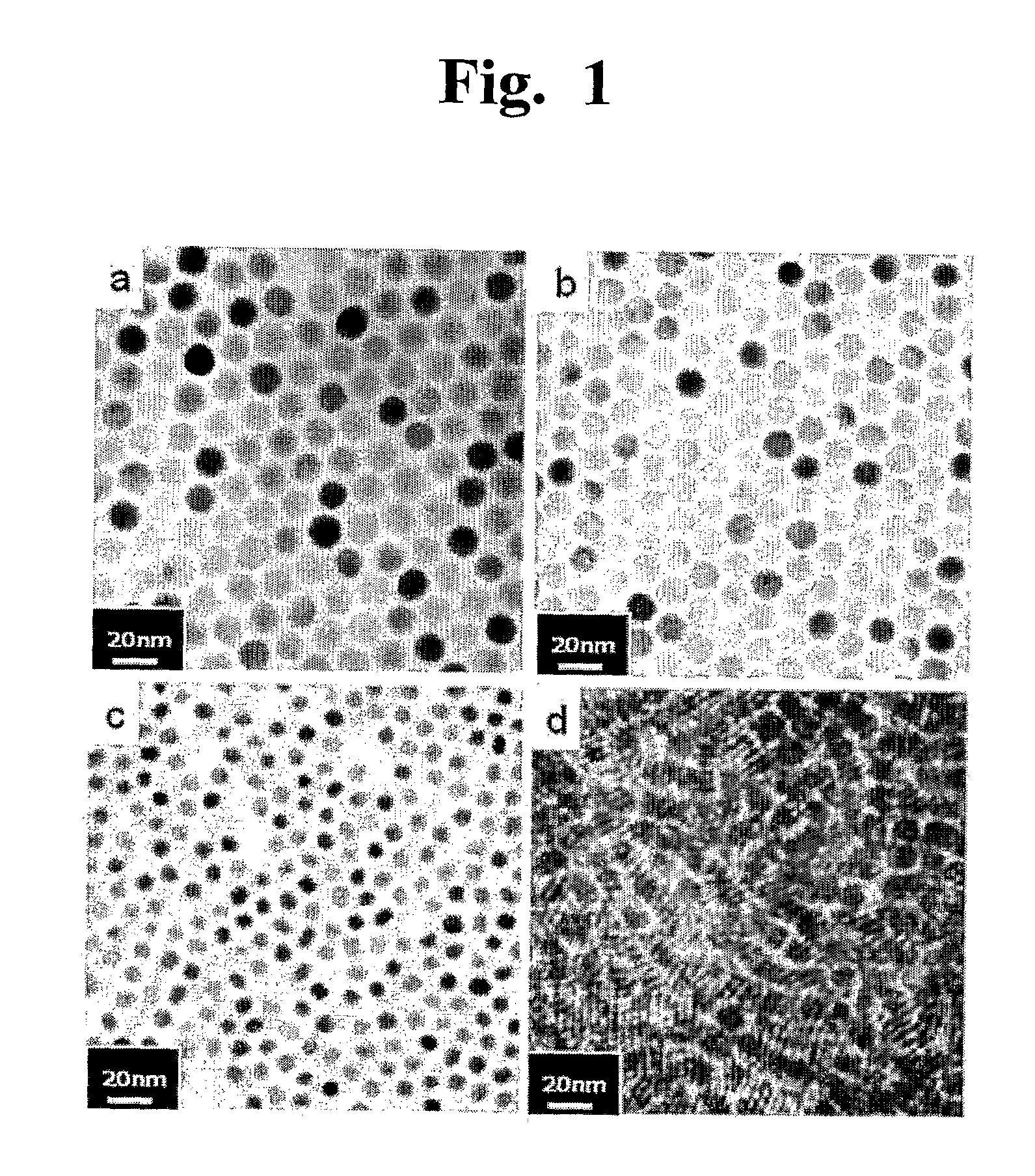
[0092]Fe3O4 and MnFe2O4 nanoparticles used in the experiments were synthesized according to the methods disclosed in Korean Pat. No. 0604975 and PCT / KR2004 / 003088. As precursors of nanoparticles, MCl2 (M=Mn2+, Fe2+, Gd2+) (Aldrich, USA) and Fe(acac)3 (Aldrich, USA), were added to trioctylamine solvent (Aldrich, USA) containing 4 mmol oleic acid (Aldrich, USA) and 4 mmol oleylamine (Aldrich, USA) as capping molecules. The mixture was incubated at 200° C. under argon gas atmosphere and further reacted at 300° C. The nanoparticles synthesized were precipitated by excess ethanol and then isolated. The isolated nanoparticles were again dispersed in toluene, generating a colloid solution. All synthetic nanoparticles exhibited a homogeneous size distribution (sa, FIG. 1b and FIG. 1d).

[0093]FePt nanoparticles used in the experiments were synthesized according to the methods known to those skilled in the art (Shouheng Sun et al. Journal of the American Chem...

example 2

Preparation of Serum Albumin-Coated Nanoparticles

[0094]Serum albumin (SA)-coated nanoparticles were prepared according to the methods described in Korean Pat. No. 10-0604975, No. 10-0652251, No. 10-0713745, PCT / KR2004 / 002509 and PCT / KR2007 / 001001. Water-insoluble nanoparticles (5 mg) obtained were dispersed in 1 mL of 1 M NMe4OH butanol solution and then homogeneously mixed for 5 min. Dark brown precipitates formed were separated by centrifugation (2,000 rpm, room temperature, 5 min). 10 mg of serum albumin (Aldrich, USA) was dissolved in 1 mL of deionized water and mixed with the precipitates, synthesizing nanoparticles coated with SA of rat. Finally, non-reactive SA was removed using a Sephacryl S-300 column (GE healthcare, USA), obtaining pure SA-coated water-soluble nanoparticles.

example 3

Preparation of Immunoglobulin G-Coated Nanoparticles

[0095]Immunoglobulin G (IgG)-coated nanoparticles were prepared according to the methods described in Korean Pat. No. 10-0604975, No. 10-0652251, No. 10-0713745, PCT / KR2004 / 002509 and PCT / KR2007 / 001001. Water-insoluble nanoparticles (5 mg) obtained were dispersed in 1 mL of 1 M NMe4OH butanol solution and then homogeneously mixed for 5 min. Dark brown precipitates formed were separated by centrifugation (2,000 rpm, room temperature, 5 min). 10 mg of human IgG (hIgG) was dissolved in 1 mL of deionized water and mixed with the precipitates, synthesizing hIgG-coated nanoparticles. Finally, non-reactive hIgG was removed using a Sephacryl S-300 column, obtaining pure hIgG-coated water-soluble nanoparticles.


PUM

PropertyMeasurementUnit
Solubility (mass)aaaaaaaaaa
Magnetizationaaaaaaaaaa
Magnetismaaaaaaaaaa

Abstract

The present invention relates a dual-modality PET (positron emission tomography)/MRI (magnetic resonance imaging) contrast agent, a hybrid nanoparticle comprising: (a) a magnetic signal generating core; (b) a water-soluble multi-functional ligand coated on the signal generating core; and (c) a positron emitting factor linked to the water-soluble multi-functional ligand. The contrast agent of the present invention is the dual-modality contrast agent enabling to perform PET and MR imaging and can effectively obtain images having the merits of PET (excellent sensitivity and high temporal resolution) and MR (high spatial resolution and anatomical information) imaging. The contrast agent of the present invention is very useful for non-invasive and highly sensitive real-time fault-free imaging of various biological events such as cell migration, diagnosis of various diseases (e.g., cancer diagnosis) and drug delivery.

Description

BACKGROUND OF THE INVENTION[0001]1. Field of the Invention[0002]The present invention relates to a dual-modality PET (positron emission tomography) / MRI (magnetic resonance imaging) contrast agent.[0003]2. Description of the Related Art[0004]As the imaging technique of biological targets becomes an increasingly important tool towards understanding of basic biological phenomena and fault-free diagnosis of various diseases, which needs to be excellent in the view of 1) sensitivity, 2) accuracy and 3) rapidity.[0005]However, current single imaging modality methods tend to be not adequate, resulting in false diagnosis. Therefore, multi-modal imaging in which each single imaging modality method is combined rapidly becomes an essential tool in the art of imaging research and a standard practice in the clinic. By using dual- or triple-modality methods, many shortcomings that are present in single imaging modality methods can be overcome. For example, several combinations of different imagin...

Claims


Application Information

Patent Timeline
IPC IPC(8): A61K51/12
CPCA61K49/0002A61K49/1854A61K49/1857A61K51/1255A61K49/1863A61K49/1869A61K49/1875A61K49/186A61K49/00A61K49/06A61K49/08
Inventor CHEON, JIN WOOCHOI, JIN-SILYOO, JEONGSOOPARK, JEONG CHANCHANG, YONGMIN
Owner IND ACADEMIC CORP FOUND YONSEI UNIV
Why Eureka
Try Eureka