Immunoliposome-nucleic acid amplification (ILNAA) assay

a technology of immunooliposome and nucleic acid, applied in the field of new assay systems, can solve the problems of reducing the likelihood of death or injury due to exposure, requiring examination of trace forensic evidence for involving biological toxins, and each of these techniques suffers from drawbacks and problems, so as to facilitate the incorporation of integral membrane proteins, facilitate the selection of attachment strategies, and enhance the range of attachment strategies.

Inactive Publication Date: 2005-07-21
UNITED STATES OF AMERICA THE AS REPRESENTED BY THE SEC OF THE ARMY
View PDF22 Cites 25 Cited by
  • Summary
  • Abstract
  • Description
  • Claims
  • Application Information

AI Technical Summary

Benefits of technology

[0040] The present invention facilitates incorporating integral membrane proteins directly into the liposomes used to encapsulate the amplification substrates. Covalent attachment of antibodies to DNA is limited to the natural phosphate group or requires the costly introduction of modified nucleotide bases into the amplification substrate through sequence-specific DNA synthesis, as for example, in streptavidin-biotin procedures which require the costly introduction of biotin into the amplification substrate.
[0041] Contrarily, in the method of the present invention, the antibody (or other specific receptor molecule) of interest is attached to the extravesicular surface of the liposome. The present invention, at once, enhances the range of attachment strategy choices thereby permitting selection of simpler and less costly attachment strategies. For example, an antibody (or other specific receptor molecule) of interest can be securely anchored to the extravesicular surface of the liposome through covalent attachment to a variety of long-chain-length hydrocarbons (12-24 carbons) that can partition into the hydrophobic core of the bilayer. Long-chain-length carboxylic acids, amines, thiols, alcohols, aldehydes, nitrites, amides, and halides are usable as such hydrocarbon anchors. Another approach involves covalently attaching the antibody to glycolipids and phospholipids, such as phosphatidylethanolamine or phosphatidylserine, through their functional groups.

Problems solved by technology

Foremost, early detection of the use by, for example, terrorists, of biological toxins will allow time for countermeasures, thus decreasing the likelihood of death or injury due to exposure.
Finally, terrorist acts involving biological toxins will frequently require examination of trace forensic evidence for the presence of toxins.
Each of these techniques suffers from drawbacks and problems.
However, even under ideal circumstances, ELISA's suffer from detection limits, which are restricted to the nanomolar (nM) to femtomolar (fM) concentration range.
The signal amplification methods employed are the same as those for other immunoassays and thus the detection limit is limited to the picomolar level with practical detection limits in the micromolar (10−6M) to nanomolar range (Wang et al., 2001; Cheng et al., 2001; Tanabe et al., 2001; Deng et al., 2001).
Taken as a whole, the immunoassay methods offer outstanding selectivity due to the specificity of the antigen-antibody interaction, but offer only modest sensitivity that is limited in practice to the nanomolar to picomolar concentration range.
However, it is not currently possible to uniquely identify a protein, such as a biological toxin, through its mono-ionic species in the MALDI-TOF mass spectrum.
Unfortunately, the kinetics of the proteolytic digestion step limits the lowest practical protein concentration to the micromolar range (Doucette et al., 2000).
Despite these advances, mass spectrometry clearly lacks both the specificity and sensitivity required for a high sensitivity assay system to detect proteins at the sub-attomolar concentration level.
The most sensitive biological signal amplification scheme ever developed, polymerase chain reaction (PCR), is limited to nucleic acid amplification and cannot directly be used to detect proteins.
Immuno-PCR methods present several difficulties, particularly for field application, as for example, in the military.
This severely limits the ability of these methods to detect very low copy numbers of antigens (10-100) as detection of only a few copies of the target DNA molecule by PCR is often difficult or impossible.
Furthermore, particularly when in the field, contamination of samples with extraneous DNA is a critical concern for samples with low target DNA concentrations.
Finally, even where amplification is successful, it entails a large and time-consuming number of amplification cycles to produce enough DNA to allow for reliable detection of the amplified product.
Although highly sensitive and specific, this method requires complex nanofabrication techniques and employs sensitive instrumentation not compatible with use in the field.
Although both techniques can detect antigens down to the level of 10-100 molecules, both require sophisticated nanofabrication and detection techniques beyond the capability of most laboratories and researchers.
In summary, none of the current cutting-edge methodologies discussed above are well suited to the development of a highly selective ultra-sensitive assay system for detecting biological toxin hazards in military deployment situations.
In the current immuno-PCR methods the amplification substrates are exposed to the bulk assay solution making DNase reduction of background DNA impossible as the amplification substrates themselves would also be degraded.

Method used


Image

  • Immunoliposome-nucleic acid amplification (ILNAA) assay
  • Immunoliposome-nucleic acid amplification (ILNAA) assay
  • Immunoliposome-nucleic acid amplification (ILNAA) assay

Examples


Embodiment Construction

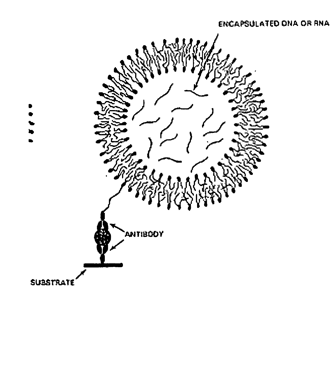
[0180] The following recites a specific example of using the invention in connection with the detection of Cholera toxin using real-time PCR for quantifying the amount of toxin present. In this case, a monosialoganglioside (GM1) a ganglioside specific for Cholera toxin, was used as the specific receptor. The immunoliposomes were formed to encapsulate an amplification substrate, which in this case, comprised 50 copies of an 85 base-pair gene fragment for human β2-microglobin. This gene fragment (amplicon) serves as the detection agent to quantify the amount of Cholera toxin present in the test sample when amplified by real-time PCR. Notably, the human B2-microglobin gene fragment is chosen strictly for convenience. Any other amplicon can be used and incorporated into the immunoliposomes using this method.

Formation of Immunoliposomes

[0181] 1) To a volume of 2 mL of chloroform is added 40 mg of 1,2-dioleoyl-sn-glycero-3-phosphocholine (DOPC), 4 mg of lissamine-rhodamine-1,2-dihexadec...


PUM

PropertyMeasurementUnit
diametersaaaaaaaaaa
diameteraaaaaaaaaa
pHaaaaaaaaaa

Abstract

Immunoliposomes and use thereof in highly specific and sensitive nucleic acid amplification assays relying on amplification of specific nucleic acid sequences released from encapsulation within a liposome after a receptor on the liposome couples with a targeted analyte/antigen immobilized on a select surface. The immunoliposome nucleic acid amplification assay permits both quantitative and qualitative analyte detection.

Description

I. FIELD OF THE INVENTION [0001] This invention relates to a new assay system and method for detecting extremely small quantities (as few as 10-1000 molecules) of compounds for which specific receptors, such as antibodies, exist. The invention is referred to as an immunoliposome-nucleic acid amplification (ILNAA) assay method. II. BACKGROUND OF THE INVENTION [0002] The early and rapid detection of biological toxins is critically important to the protection of security personnel deployed in hostile situations or in instances of domestic terrorism. Biological toxins, such as botulinum toxin, are lethal at very low concentrations, which necessitates detection measures that are both highly specific and extremely sensitive. There are a multitude of scenarios that may require the ability to detect biological toxins at sub-attomolar (10−18M) concentrations or even at levels approaching a few molecules. Foremost, early detection of the use by, for example, terrorists, of biological toxins w...

Claims


Application Information

Patent Timeline
Patent Type & Authority Applications(United States)
IPC IPC(8): A61K9/127A61K48/00C12N15/88C12P19/34C12Q1/68
CPCA61K9/127C12Q1/6806C12Q2563/161
Inventor O'LEARY, TIMOTHYMASON, JEFFREY
Owner UNITED STATES OF AMERICA THE AS REPRESENTED BY THE SEC OF THE ARMY
Why Eureka
Try Eureka