Ultrasound interfacing device for tissue imaging

Active Publication Date: 2005-10-27
ISCI SURGICAL
View PDF42 Cites 160 Cited by
  • Summary
  • Abstract
  • Description
  • Claims
  • Application Information

AI Technical Summary

Benefits of technology

[0024] The present invention is a device to acoustically couple a high frequency ultrasound transducer to the surface of the tissues to be imaged. The device provides an acoustic pathway of minimal effect on the ultrasound signals. The device may be directly placed on the tissue region to be imaged to act as a standoff and aid in placing the desired tissue region into the focal zone of the ultrasound

Problems solved by technology

The resolution of ultrasound imaging is inherently limited by the frequency of the ultrasound used, with higher frequency providing higher resolution but at the expense of reduced tissue penetration.
However with high frequencies, it is technically difficult to fabricate a phased array of transducer elements due to the small wavelengths and resultant precision and small size required of the array.
In ranges at and above 50 MHz, most commonly used coupling materia

Method used


Image

  • Ultrasound interfacing device for tissue imaging
  • Ultrasound interfacing device for tissue imaging
  • Ultrasound interfacing device for tissue imaging

Examples


example 1

Fabrication and Testing of Ultrasound Interfacing Devices

[0043] Various materials were prepared as films and investigated to determine suitability for use as a scan window for high frequency ultrasound imaging. Each material sample was fixed in a machined Delrin cap and attached to the scan head of a high frequency ultrasound handpiece. The ultrasound imaging system was set up with a flat plate target at the focal distance of the ultrasound transducer. The system operated at a center frequency from 50-70 MHz. The samples were imaged under the same conditions and were evaluated for surface reflectivity, reverberations, and attenuation of the ultrasound signal. The following table lists the materials and conditions.

MaterialConditionUrethane / Silicone Copolymer - Elasteon, AortechShore 80A, 125 micron thick filmIntlSilicone RTV - General ElectricCast film from solvent dispersed RTV,˜650 micron thickSilicone RTV-L Silastic - Dow CorningCast film from solvent dispersed RTV,˜650 micron ...

example 2

Ultrasound Characterization of Scan Window Material Candidates

[0045] Ultrasound signal loss and time-of-flight characteristics were measured on candidate scan-window polymer samples. These quantities were measured with respect to a 2-way flat plate impulse response with a water-only path.

[0046] The test system for measuring the signal loss consisted of: a Tektronix TDS 544A oscilloscope, Panametrics 5910R Pulser-receiver, a high frequency ultrasound transducer with a 62 MHz center frequency, 6 mm focal length and approximately 45% bandwidth, and a fused silica flat plate target. All measurements were taken with 50 dB of attenuation on the Panametrics receiver. The samples were placed at a distance approximating where they would be if used as a scan window located 4 mm away from the transducer face or 2 mm away from the nominal focus. An impulse response was first maximized without any sample by obtaining peak signal amplitude by varying the depth and target angle. The 64× average ...

example 3

Sterilized Ultrasound Interfacing Device

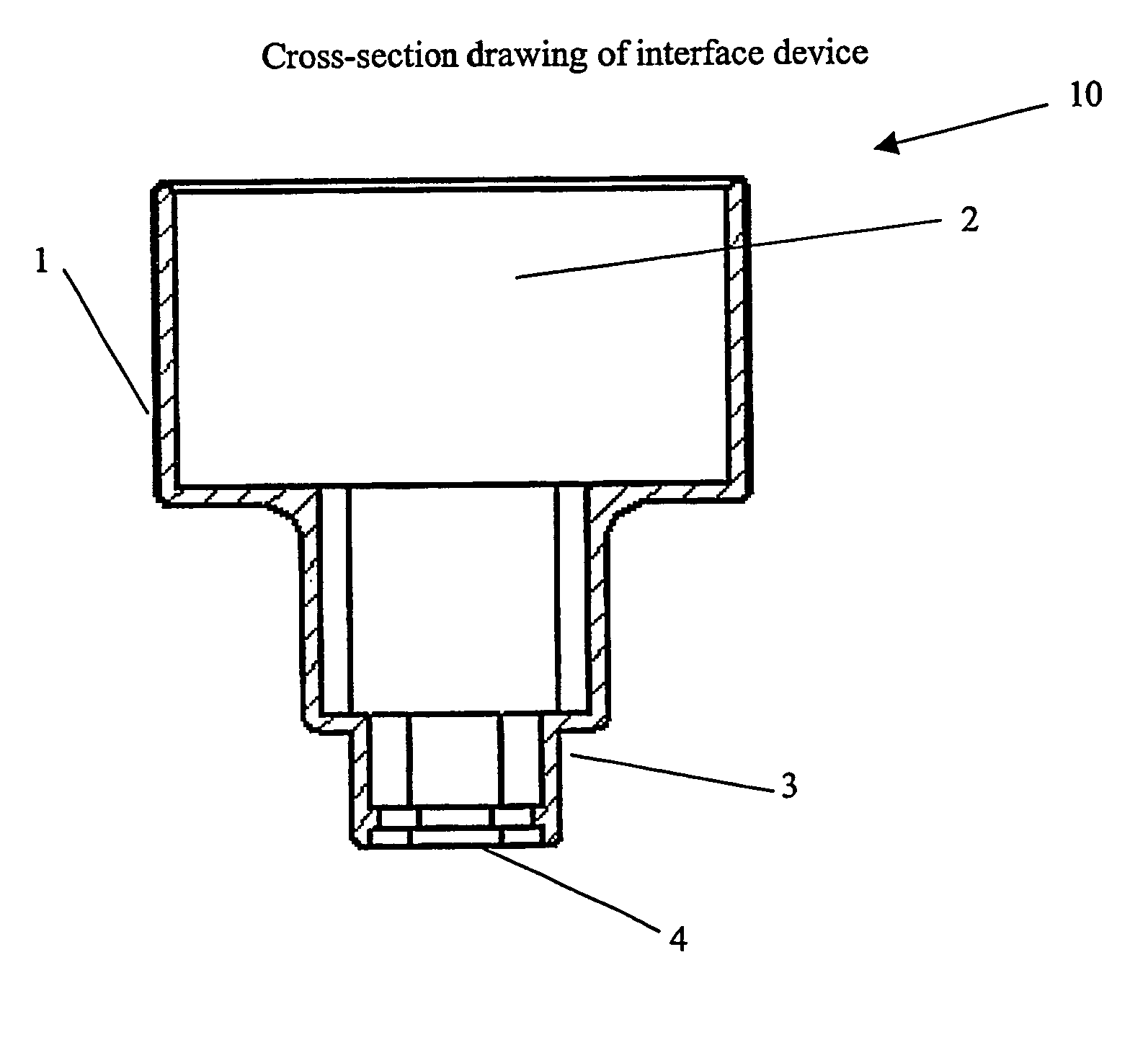
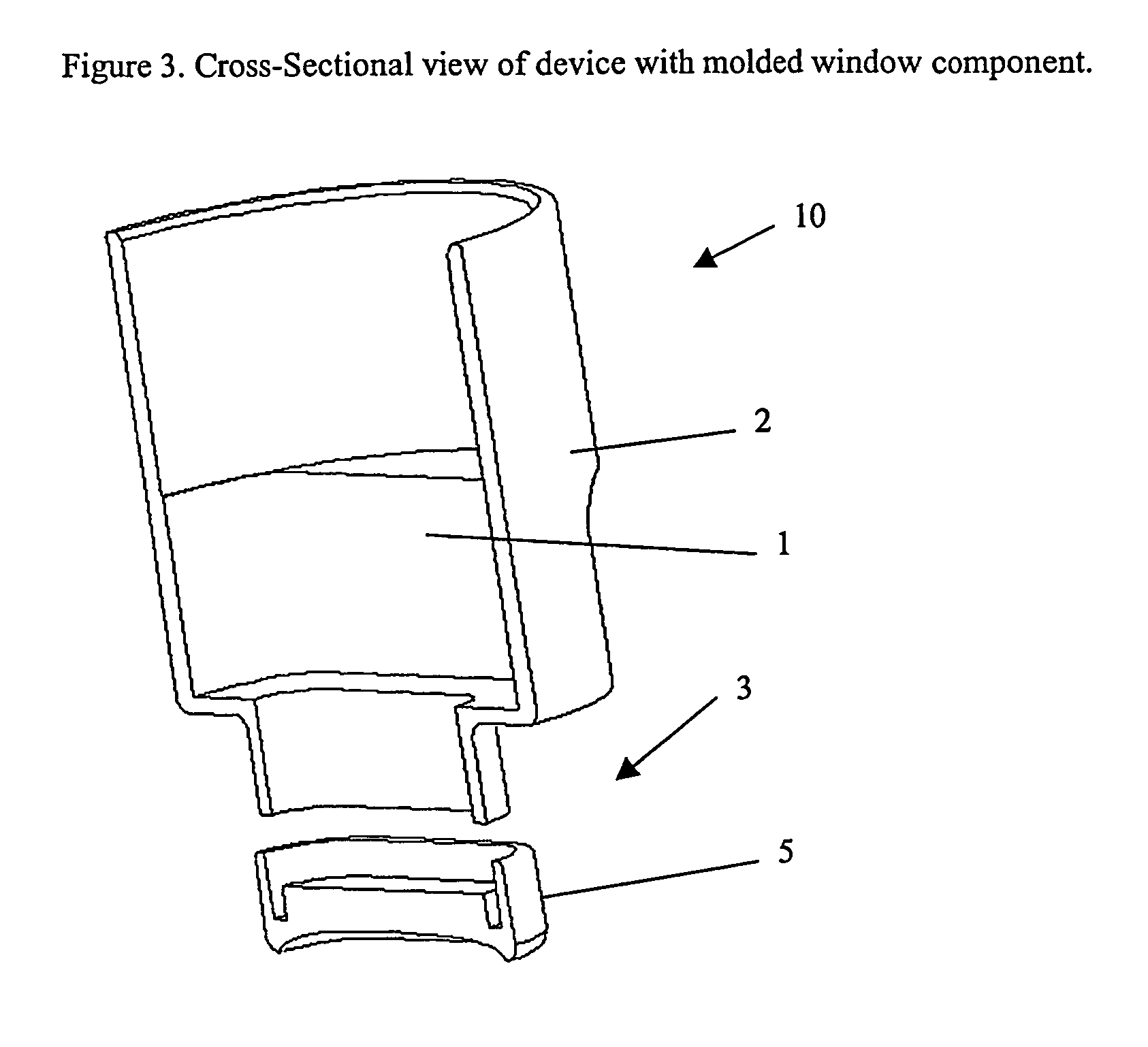
[0050] Ultrasound interfacing devices were fabricated for use with the high frequency ultrasound imaging system described above. The devices were formed of machined nylon 66 caps designed to attach to the distal end of the scanhead. The distal end of the device was a rectangular window opening for the imaging hydrogel, the opening being 4 mm×9 mm in dimension. The distal segment of the device was designed to provide for mechanical entrapment of the gel by use of contours on the interior surface.

[0051] Polyurethane hydrogel scan windows were cast into the devices in varying polymer concentrations and used for imaging on ex-vivo and in-vivo human eyes. The polyisocyanate prepolymer was diluted with acetonitrile to concentrations of 20%, 15% and 10% by weight. A solution of 50 mM sodium bicarbonate was prepared and adjusted to a pH in the range of 7.6-7.8. The prepolymer solutions were mixed with the bicarbonate solutions in a 1:1 ratio to yiel...


PUM


Abstract

A device (10) used with high frequency ultrasound provides a replaceable barrier between the ultrasound transducer and the tissues being imaged. The device (10) provides an acoustic pathway of minimal effect on the ultrasound signals, and furthermore provides for a safety barrier between a mechanical scanner and the tissues being imaged. The configuration of the device (10) allows it to function as a standoff for the ultrasound transducer to place the focus of the ultrasound beam at the desired depth of tissue for imaging. In addition, the device (10) may be sterilized and replaced as needed, providing sterile tissue contact surfaces. The device (10) has additional features, which are especially advantageous for diagnosis and surgery of the eye.

Description

BACKGROUND OF INVENTION [0001] Ultrasound imaging is routinely used in medicine for non-invasive imaging of tissues and organs, both for diagnosis and for image guided surgery. The resolution of ultrasound imaging is inherently limited by the frequency of the ultrasound used, with higher frequency providing higher resolution but at the expense of reduced tissue penetration. The most common forms of medical ultrasound imaging, cardiac and fetal imaging, utilize ultrasound frequencies in the range of approximately 2 to 10 Megahertz (MHz) in order to image deep tissues. The ultrasound transducers used in general imaging are typically phased array designs, with a plurality of fixed transducer elements used to form a scan head of a probe. The individual trnsducer elements are switched electronically to sweep the ultrasound beam across an area to be imaged at high frame rates. [0002] High frequency ultrasound imaging, generally in the range of 50 to 100 MHz, is used to obtain images of sm...

Claims


Application Information

Patent Timeline
IPC IPC(8): A61B8/00A61B8/10A61B8/12A61B3/10A61B8/14
CPCA61B8/10A61B8/4422A61B8/4281
Inventor RACHLIN, DANIELCONSTON, STANLEY R.
Owner ISCI SURGICAL
Why Eureka
Try Eureka