
Method and apparatus for inhibiting the growth of and shrinking cancerous tumors

a cancerous tumor and tumor growth technology, applied in the field of methods and apparatus for inhibiting the growth and shrinking of cancerous tumors, can solve the problems of reducing the desirability of implants, increasing the likelihood of complications associated with procedures, and invasive techniques, so as to reduce the cost of surgery, many people are unable to have their abnormalities corrected or enhanced, and the effect of reducing the likelihood of complications

Inactive Publication Date: 2005-11-03
BRAVA
View PDF66 Cites 41 Cited by
  • Summary
  • Abstract
  • Description
  • Claims
  • Application Information

AI Technical Summary

Benefits of technology

[0008] Each of these above-mentioned apparatuses and methods requires an invasive surgical technique to accomplish the soft tissue expansion. Invasive techniques increase the likelihood of the complications associated with the procedure including those mentioned above with respect to implant surgery. In addition, the expense of surgery precludes many persons from having their abnormalities corrected or physical attributes enhanced.
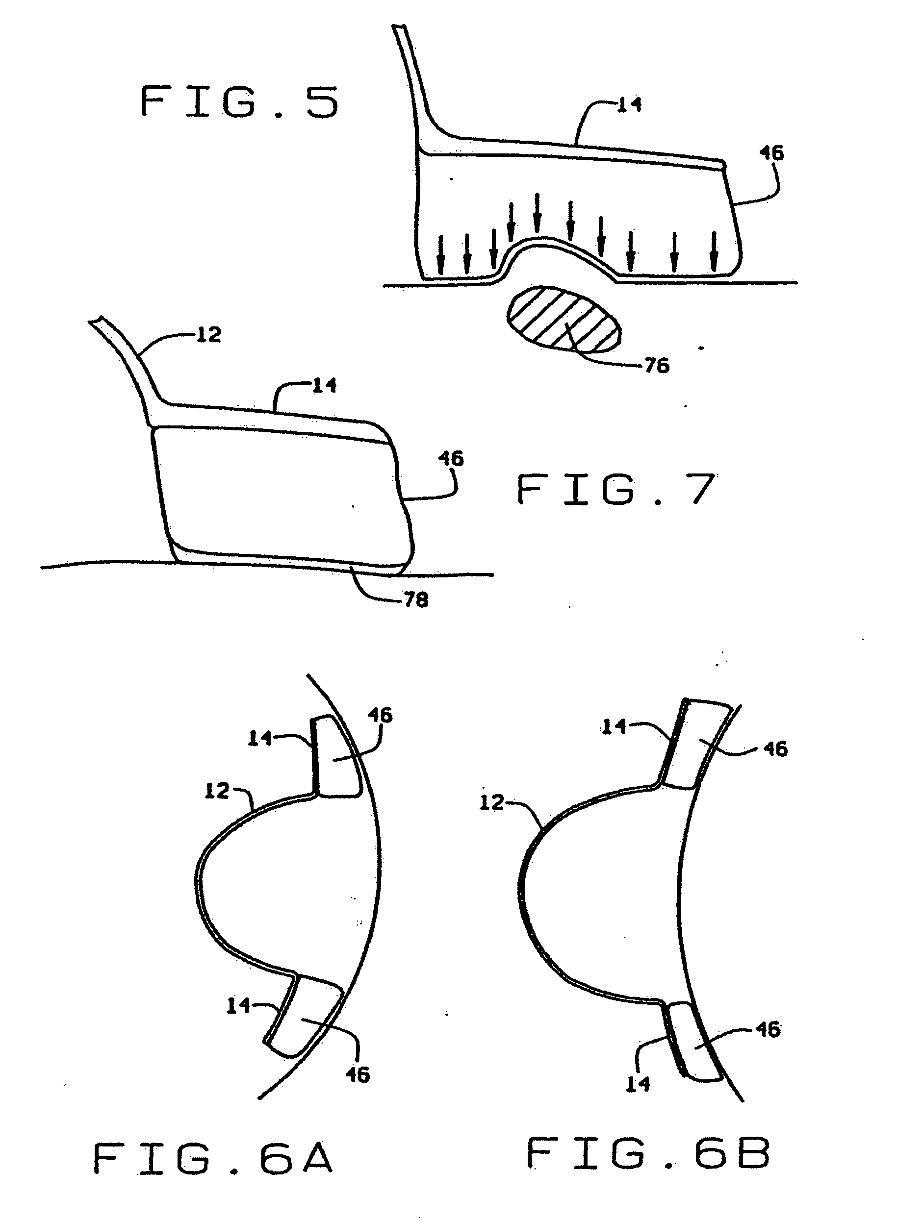
[0012] Most of these prior art devices and methods have failed to achieve long term soft tissue enlargement while preventing damage to the soft tissue being enlarged, as well as surrounding tissue. As disclosed and claimed in the parent applications and patents noted above, the inventor herein has succeeded in designing and developing a new generalized method and apparatus for soft tissue enlargement which prevents damage to soft tissue. The apparatus used for this enlargement is comprised of a rigid, fluid-impervious dome having a rim about its periphery and a vacuum pump for reducing pressure within the dome. The rim has sufficient surface area such that the pressure applied to the patient by the rim is less than or equal to the negative pressure applied to the soft tissue under the dome. In the parent patent filings, one specific teaching to achieve this balanced force utilized a rim with substantially the same cross-sectional area as the normal area of the dome. Thus, as long as pressure within the dome is regulated to a limit below which medical complications will not occur, the opposing contact pressure against the patient is below this threshold as well. With this approach, damage is avoided not only to the soft tissue being enlarged, but the surrounding tissue as well. In the preferred embodiment of the apparatus, the vacuum pump has a self-contained power source. In addition, a pressure sensor and servomechanism control the pump such that the vacuum within the dome is maintained at a magnitude less than 35 mmHg. Variant embodiments may be configured to fit over and enlarge a human breast, or any other desired soft tissue area.
[0013] In still another patent issued to one of the present inventors entitled “Method And Apparatus For Promoting Soft Tissue Enlargement and Wound Healing”, U.S. Pat. No. 5,701,917, issued Dec. 30, 1997, the disclosure of which is incorporated by reference, an invention was disclosed which utilizes a rigid fluid-impervious dome having a rim about its periphery and a vacuum pump for reducing pressure to thereby apply a distracting force to the soft tissue isolated by and within the dome. The dome may be conveniently located over an open wound in order to promote healing of the wound by enlarging the soft tissue under the dome. As the soft tissue grows, it promotes healing of the wound through acceleration of the closing thereof by soft tissue growth. As wounds may be received by a patient to any part of his body, the co-inventor's prior disclosed and claimed invention includes the use of a dome over virtually any part of the human body.
[0015] It is well recognized in the medical literature that decubitus ulcers are caused by unrelieved external pressure that occludes blood flow and results in tissue necrosis. In recognition of this fact, these ulcers are called pressure sores. The average capillary pressure in human skin is around 15-20 mmHg. E. M. Landis, Micro-Injection Studies of Capillary Blood Pressure in Human Skin, 15 Heart 209-228, (1930). For convenience, 20 mmHg has been used to describe this pressure throughout the previous patent filings. However, it should be understood that pressures below 20 mmHg may also be used without departing from the scope of these inventions and that these lower pressures may provide additional margins in preventing damage to tissues. Therefore, the local application of an external pressure up to 20 mmHg will not collapse capillaries adjacent the location of the applied pressure and thus will not disturb the circulation. Therefore, local application of contact pressures less than or equal to 20 mmHg are well tolerated for prolonged periods of time. This tolerance has been confirmed by the inventor through use of a prototype which did not cause adverse effects after many hours of continuous use as long as the pressure under the rim remained below or around 20 mmHg.
[0018] These findings are consistent with the clinical testing of the prototype of the breast device. It was found that a continuous pressure under the rim of 40 mmHg could be tolerated for only one hour by healthy volunteers. After one hour, the volunteers started to complain of pain which is the warning sign of impending tissue damage. Higher pressures led to pain under the rim after even shorter periods of time. Lower pressures around 30 mmHg led to pain after 4 hours. However, if the pressure is allowed to cycle, that is if it is dropped down to 0-20 mmHg to allow the tissues to temporarily reperfuse for a few minutes, higher peak pressures can be tolerated. The higher the peak pressures, the shorter they are tolerated and the longer the low pressure part of the cycle needs to be to allow the tissues to recuperate.
[0021] As indicated by the summary of the medical literature given above, a vacuum dome may also be used in alternative methods in keeping within the scope of the inventor's concept. For example, the device might have a rim cross-sectional area substantially less than the normal area of the dome and be used in either of two methods. In a first method, a somewhat lower vacuum pressure may be induced in the dome such that the opposing contact pressure under the rim may be maintained at bearable pressures for extended periods of time and yet provide a therapeutic effect. Alternatively, the vacuum in the dome may be regulated in a routine which provides somewhat higher vacuum pressures in the dome for shortened periods of time separated by periods of lower vacuum pressures to allow tissue reperfusion. In other words, alternating cycles of high vacuum, tissue reperfusion, high vacuum, tissue reperfusion, etc., may achieve a therapeutic effect in enlarging the soft tissues. With either of these methods, the rim may have a cross-sectional area substantially less than the normal area of the dome.

Problems solved by technology

However, the severity of the potential complications including scarring, implant rupture, capsular contracture, necrosis and implant migration as well as the recent adverse publicity thereof have significantly reduced the desirability of these implants.
Invasive techniques increase the likelihood of the complications associated with the procedure including those mentioned above with respect to implant surgery.
In addition, the expense of surgery precludes many persons from having their abnormalities corrected or physical attributes enhanced.
Although this device is sufficient for its intended purpose, it is not suitable for general soft tissue enlargement.
Laceration and contusion can occur if too strong of a suction is applied to soft tissue.
As the pressure within the inverted nipple instrument is not regulated, contusion or laceration can occur.
Excessive contact forces against the patient can cause ulceration, laceration, and contusions.
As the contact forces are not regulated in the nipple instrument, these further complications also can occur.
In addition, general soft tissue enlargement is not feasible with the instrument due to the size and shape of the cup.
However, this patent provides no teaching as to the pressures to be used, the potential danger to the skin tissues, or any suggestions as to how the device is to be retained in place during use.
Apparently, the device is used in a clinical setting and is not suitable for long term wear such as for 8-10 hours.
This patent has been the subject of ridicule by at least one medical authority.
Most of these prior art devices and methods have failed to achieve long term soft tissue enlargement while preventing damage to the soft tissue being enlarged, as well as surrounding tissue.
Tissues can tolerate short periods of ischemia, but if the pressure is continuous and perfusion is not restored within a relatively short period of time, tissue damage will ordinarily ensue.
For instance, 20 mmHg is well tolerated for prolonged periods of time, but 40 mmHg will lead to tissue injury if the pressure is not relieved for 13 hours.
After one hour, the volunteers started to complain of pain which is the warning sign of impending tissue damage.
Higher pressures led to pain under the rim after even shorter periods of time.
Lower pressures around 30 mmHg led to pain after 4 hours.
Therefore, pressures under the rim greater than 20 mmHg can only be tolerated if there is a means to continuously cycle the pressure peaks on and off allowing for tissue re-perfusion during the off periods.
In utilizing this construction, the inventor is aware of potential complications which can develop when an area of the body needs to be enclosed for prolonged periods of time within the dome having an airtight seal.
For example, while a rim made of conforming or other soft materials may suffice for temporary use, a number of problems arise in the skin contact area when prolonged negative pressure application is necessary.
Thus, admittedly, the pool of users did not totally reflect the female population as a whole.
Yet more than three decades of experience on millions of women leave no evidence that expanding that residual cancer prone breast increases the likelihood of cancer growth or recurrence.
Furthermore, despite the fact that skin is the most cancer prone organ in the body and skin expansion is routinely used in plastic surgery, there are no reports in the literature of cancer arising in the expanded skin.

Method used


Image

  • Method and apparatus for inhibiting the growth of and shrinking cancerous tumors
  • Method and apparatus for inhibiting the growth of and shrinking cancerous tumors
  • Method and apparatus for inhibiting the growth of and shrinking cancerous tumors

Examples


Embodiment Construction

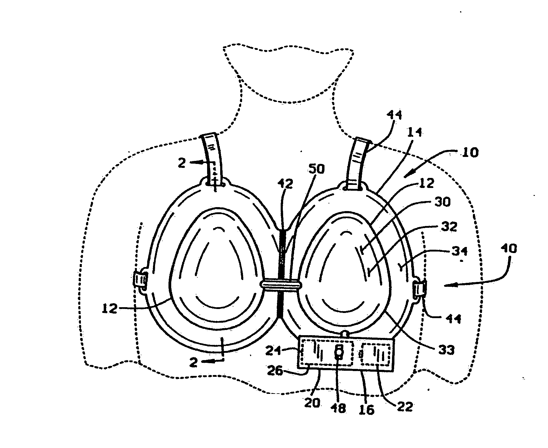
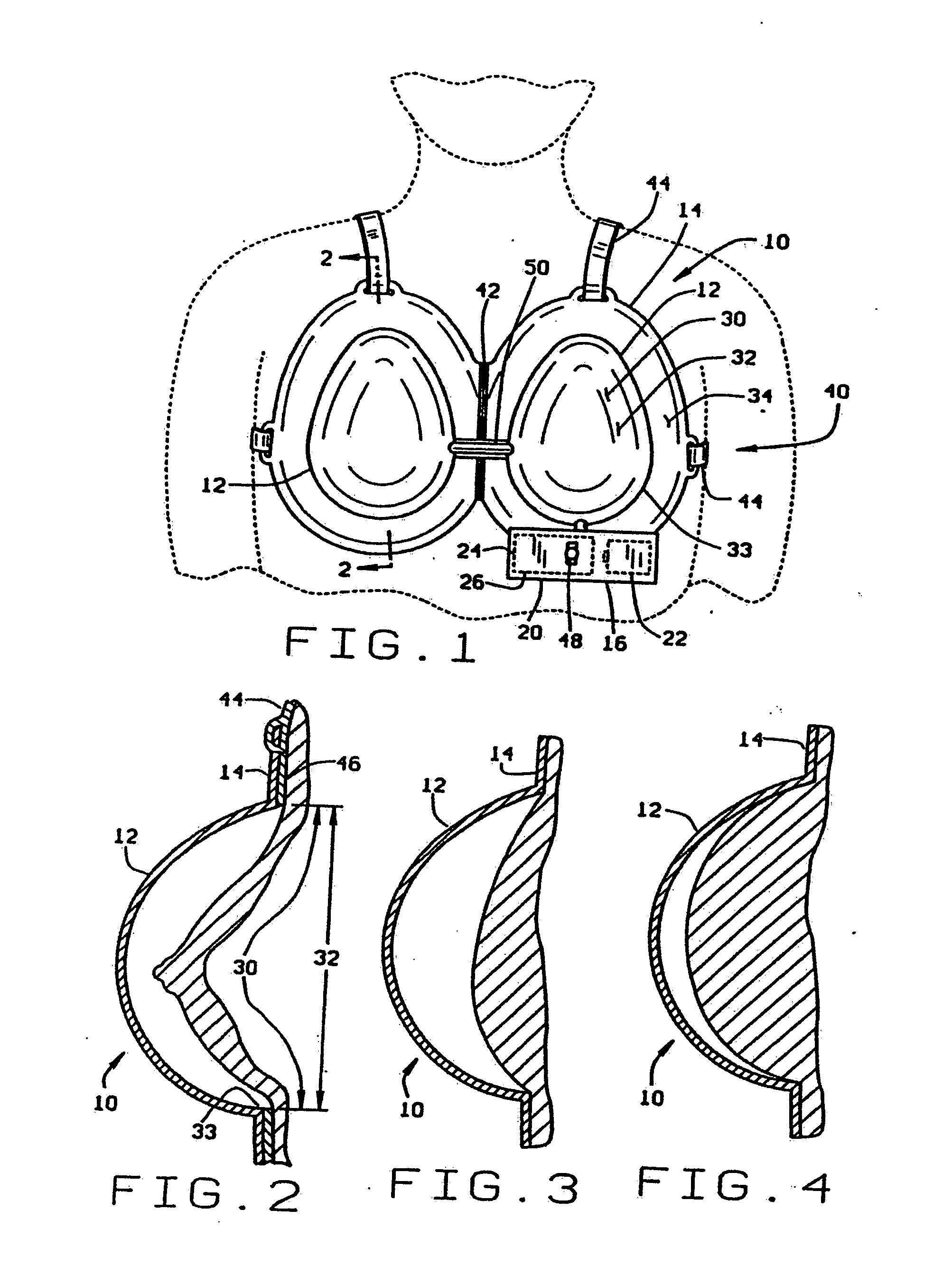
[0051] One embodiment of the soft tissue enlargement apparatus 10 is generally comprised of a dome 12 having a rim 14 and a vacuum pump assembly 16 for creating a vacuum within the dome. Although the vacuum pump assembly 16 may be a separate hand-held pump in one variant embodiment, in one preferred embodiment the vacuum pump assembly 16 is a self-contained vacuum pump 20 with an independent power source 22, pressure sensor 24, and servomechanism 26 for driving, regulating and controlling the vacuum pump 20.

[0052] Regulation of the vacuum within the dome is essential to prevent contusions caused by rupturing capillaries adjacent the surface of the skin. Medical data suggest that these contusions will not occur if vacuum within the dome is maintained at less than about 20-25 mmHg, at least on a continuous basis. Thus, the vacuum pump 20 must be regulated to control the vacuum within the dome to within this limit presuming a protocol involving extended wearing at continuous pressure ...


PUM


Abstract

A dome for applying a vacuum to a patient's breast is comprised of a generally rigid dome capable of withstanding a pressure differential, with a rim and rim cushion underlying the rim of the dome for supporting a rim from the patient's skin surface. The rim may be generally wider than the dome in order to distribute the attendant forces across a greater surface and avoid tissue damage. A sticky sole underlies the rim cushion and seals the rim cushion to the patient's skin to thereby preserve the vacuum within the dome. The sticky sole may be comprised of any adhesive material or even be achieved through the use of an appropriate material for the rim cushion itself. A portable pump unit is connected to the domes and maintains the vacuum within the domes during a recommended protocol. By using the device in accordance with the prescribed protocol, the growth of cancer cells and tumors within the breasts are inhibited.

Description

[0001] This application is a continuation-in-part of co-pending U.S. patent application Ser. No. 09 / 818,812 filed Mar. 27, 2001, which is a continuation of co-pending U.S. patent application Ser. No. 09 / 141,460 filed Aug. 27, 1998 entitled “Vacuum Dome With Supporting Rim And Rim Cushion,” which is a continuation of U.S. patent application Ser. No. 08 / 698,941 filed Aug. 16, 1996 entitled “Vacuum Dome With Supporting Rim And Rim Cushion” (now abandoned), which is a continuation-in-part of U.S. patent application Ser. No. 08 / 516,623 filed Aug. 18, 1995 entitled “Method And Apparatus For Soft Tissue Enlargement With Balanced Force Appliance” (now U.S. Pat. No. 5,676,634), which is a continuation-in-part of U.S. patent application Ser. No. 08 / 504,640 filed Jul. 20, 1995 entitled “Method And Apparatus For Soft Tissue Enlargement By Distractive Force” (now U.S. Pat. No. 5,695,445), which is a continuation of U.S. patent application Ser. No. 08 / 220,186 filed Mar. 30, 1994 entitled “Method ...

Claims


Application Information

Patent Timeline
IPC IPC(8): A61B19/00A61F5/00A61F5/41A61H7/00A61H9/00A61H19/00
CPCA61F5/41A61F2005/412A61H9/005A61H2201/1621A61H2201/165A61H2205/082A61H2201/1616A61H19/00A61B90/02A61B2017/00796
Inventor FREYRE, CARLOS V.KHOURI, ROGER K.
Owner BRAVA
Learn More