
Method for identifying protein-protein interactions

a protein and interaction technology, applied in the field of protein interaction detection methods, can solve the problems of laborious process, failure to describe or suggest, and each has its own limitations, and achieve the effect of limiting background growth

Inactive Publication Date: 2006-01-26
ESBATECH
View PDF2 Cites 0 Cited by
  • Summary
  • Abstract
  • Description
  • Claims
  • Application Information

AI Technical Summary

Benefits of technology

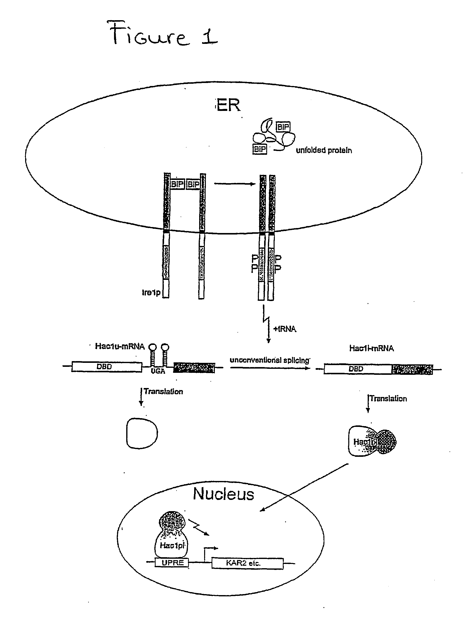
[0025] Advantages of the present invention include the ability to detect protein-protein interactions in the endoplasmic reticulum. This is an advantage because in cellular growth selection assays, all the cells in the neighborhood of a cell secreting a ligand which functionally interacts with a receptor would profit and thus grow, even if they express an unrelated ligand. Expression of the receptors and their soluble ligand in a closed compartment such as the ER, which provides the same properties as the extracellular space, should limit such background growth caused by the diffusion of the ligand.
[0026] Additionally, the use of the screening system described herein to find targets for extracellular protein-protein interactions, for example single chain antibodies, is a useful alternative to the phage display method, and provides the advantage of circumventing the need to purify target proteins. A single chain library fused to the C-terminus of Ire1p co-expressed with the fusion of a target protein to the C-terminus of Ire1p enables for the selection of proteins capable of binding the single chain antibody.

Problems solved by technology

These and other systems for identifying protein-protein interactions are useful in certain contexts, however, each has its own limitations.
However, the prior art in general and the '408 application specifically fail to describe or suggest, for example, various advantageous read-out systems.
However these methods all have drawbacks, for example, by requiring purification of the antigen.
This can be a laborious process.

Method used


Image

  • Method for identifying protein-protein interactions
  • Method for identifying protein-protein interactions
  • Method for identifying protein-protein interactions

Examples


example 1

[0244] The NLD of the Ire1 complementing mutants was substituted with known interacting partners in order to make the induction of the UPR pathway and consequent reporter gene expression dependent on a specific interaction happening either in the ER-lumen or in the cytoplasm.

[0245] Depending on the original location of the studied proteins, the respective Ire1p fusions were expressed either in the ER or in the cytoplasm.

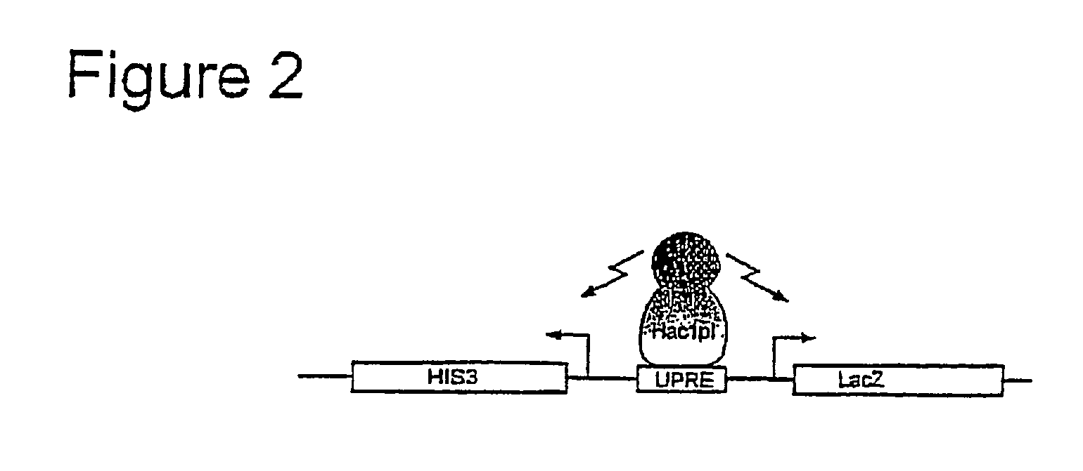
[0246] The activated Hac1pi induces expression of two selectable reporter genes, HIS3 and LacZ that bear a UPRE sequence upstream of their divergent promoters (FIG. 2).

[0247] Cloning of the Ire1 Fusions

[0248] IRE1 DNA sequences were amplified from yeast genomic DNA by PCR with proof-start polymerase (QIAGEN) using primers that contained restriction sites at their 5′ end. To generate the Ire1K702R point mutation, two additional primers harbouring a base-pair change were used to amplify a 5′ fragment and a 3′ fragment of the Ire1 C-terminus, each harbouring the res...

example 2

The Transmembrane Domain is not Necessary for the Ire1p activity

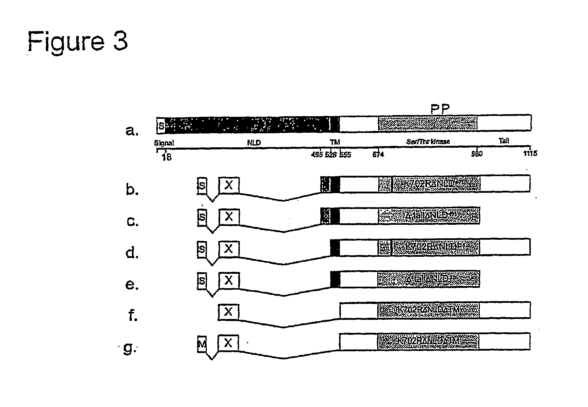
[0259] Ire1p is localized in the ER membrane and signals to the nucleus if unfolded proteins accumulate in the ER lumen. To test whether the association of Ire1p with the ER membrane is necessary for its fixation, JunLZ was fused with a Ire1p C-terminal fragment that lacks the transmembrane domain (TM) (Ire1ΔNLDΔTM). A myristoilation signal (M) was also added to the N-terminus of this fusion protein. FIG. 5 shows that, although the construct containing the MS had a higher activity then the one lacking the MS, both fusion proteins strongly activated Hac1p-dependent reporter gene expression.

[0260] Surprisingly, both Ire1 ΔNLDΔTM derivatives exhibiting dimerization ability were further activated by Tunicamycin. In contrast, the same cytoplasmic Ire1p fragment lacking a functional dimerization motif showed no constitutive activity and was also not inducible by Tunicamycin. These results indicate that, upon dimerization, ...

example 3

[0261] Ligands Bind Specifically to their Receptors in the ER Lumen

[0262] Although the growth hormone (GH) and the extracellular domain of its receptor can interact in a nuclear two-hybrid assay [11], the oxidizing environment of the secretory pathway and the extracellular matrix of living organisms can be a prerequisite for the proper folding and stability of many extracellular proteins, and might be obligatory for the function of other receptor-ligand pairs. By fusing the extracellular domains of receptors (mouse EGF receptor and mouse FLT1) to Ire1K702R526, and their specific ligands (mEGF and mVEGF) to Ire1tailΔNLD495, a system in which only co-expression of the appropriate receptor-ligand fusion protein pair should be able to activate the reporter genes was generated. In this system, potential autodimerization of ligand or receptor fusions cannot activate reporter gene expression because the receptors are fused to K702R mutants and the ligands to Δtail mutants. Binding of the ...


PUM

PropertyMeasurementUnit
Timeaaaaaaaaaa
Lengthaaaaaaaaaa
Interactionaaaaaaaaaa

Abstract

The properties of yeast help, a type I ER membrane protein which is involved in the unfolded protein response (UPR), have been exploited to develop â. system for the detection and study of interactions between extracellular and / or membrane proteins. In the system, proteins of interest are fused to the lumenal N-terminus of a truncated Ire1p. A specific interaction between two partners may be visualized through dimerization of the Ire1p moiety which, either, directly or indirectly, results in a detection means, for example, the expression of a selectable reporter gene. Depending on the type of reporter gene used, its expression can positively or negatively influence cell growth, thus allowing selection of both stimulation and inhibition of protein-protein interactions. The system presented here can also be used to study intracellular protein interactions.

Description

CROSS REFERENCE TO RELATED APPLICATIONS [0001] This application claims priority from U.S. Provisional Application No. 60 / 382,774, filed May 22, 2002.BACKGROUND OF THE INVENTION [0002] 1. Field of the Invention [0003] The present invention relates to a method for detecting the interaction of proteins using biological techniques. [0004] 2. Background of the Related Art [0005] Methods for Identifying Protein-Protein Interactions [0006] Protein-protein interactions provide the basis for critical and diverse biological functions. For example, transcription, DNA replication, enzyme regulation and assembly, antigen-antibody reactions and receptor-ligand systems all depend in some way on protein-protein interactions. It is also through protein-protein interactions that disease states and oncogenesis are perpetuated. It is, therefore, of interest to identify protein-protein interactions. [0007] In addition to using well known biochemical techniques to study protein-protein interactions, a me...

Claims


Application Information

Patent Timeline
IPC IPC(8): C12Q1/70C12Q1/68G01N33/53C12P21/06C12N9/12G01N33/567C12N15/10
CPCC07K2319/00C12N9/12C12Q1/6897C12Q1/02C12N15/1055
Inventor URECH, DAVID M.LICHTLEN, PETERBARBERIS, ALCIDE
Owner ESBATECH
Learn More