Combined MR coil technology in medical devices

a technology of medical devices and coils, applied in the field of medical devices, can solve the problems of not being able to permit imaging, occupying a large space, x-ray exposure for any extended period of time is harmful to the patient, etc., and achieves the effects of reducing scan time, reducing snr, and improving spatial and temporal mr imaging

Inactive Publication Date: 2006-04-06
NEXGEN +1
View PDF31 Cites 19 Cited by
  • Summary
  • Abstract
  • Description
  • Claims
  • Application Information

AI Technical Summary

Benefits of technology

[0017] A combination of RF microcoil(s) and surface coil(s) can be used with a preamplifier positioned on the delivery device (or distally connected by electronics or wires / cables to the combination of microcoils), which serves to amplify signals from the RF microcoils. More than one surface coil and more than one microcoil may be present, as the distribution of microcoils along a length of the image area or the medical device (e.g., a catheter) helps to define the adjacent region(s) better within which local signals are detected. The coils may add or integrate or otherwise combine their detectable volumes, thereby defining a combined volume (the term “combined volume” will be used to mean the field volumes produced by both the surface coil(s) and the microcoil(s) used during imaging) which can be efficiently observed by the MR system.
[0027] An eighth aspect of the present technology is to use MR microcoils and MR surface coils interactively and simultaneously, to eliminate the current “searchlight in the dark” problem of imaging with only a single microcoil.

Problems solved by technology

X-ray imaging, such as X-ray fluoroscopy, is a possible method of providing a view of the procedural area, but X-ray exposure for any extended period of time is itself harmful to the patient.
Fiber optic viewing of the area does not provide any harmful radiation to the patient, but the fiber optics may take up too large a space to provide both the light necessary for viewing and a path for return of the light, and does not permit imaging beyond the surface (that is, only the surfaces of internal objects may be viewed from the position where the fiber optic device is located).

Method used


Image

  • Combined MR coil technology in medical devices
  • Combined MR coil technology in medical devices
  • Combined MR coil technology in medical devices

Examples


Embodiment Construction

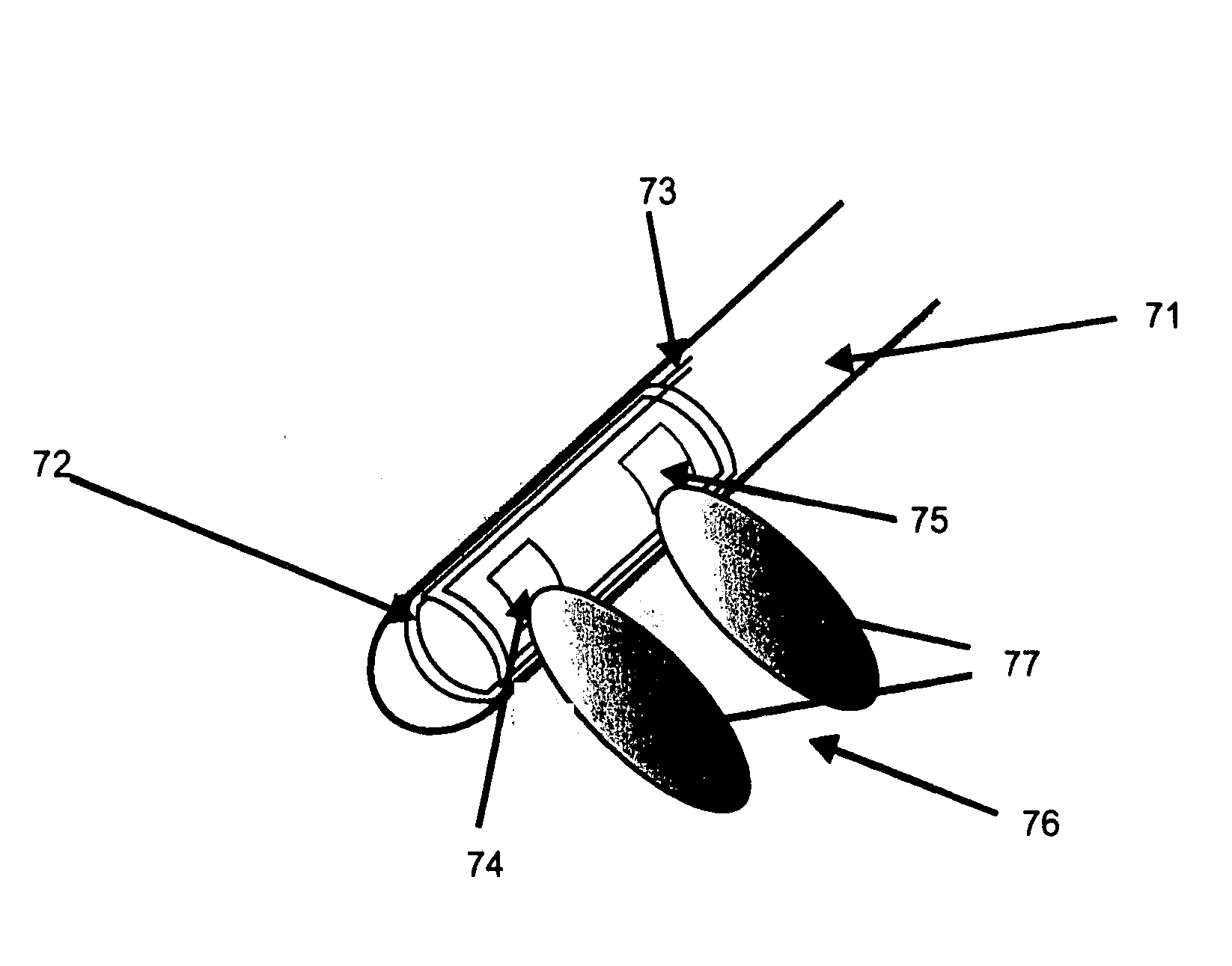
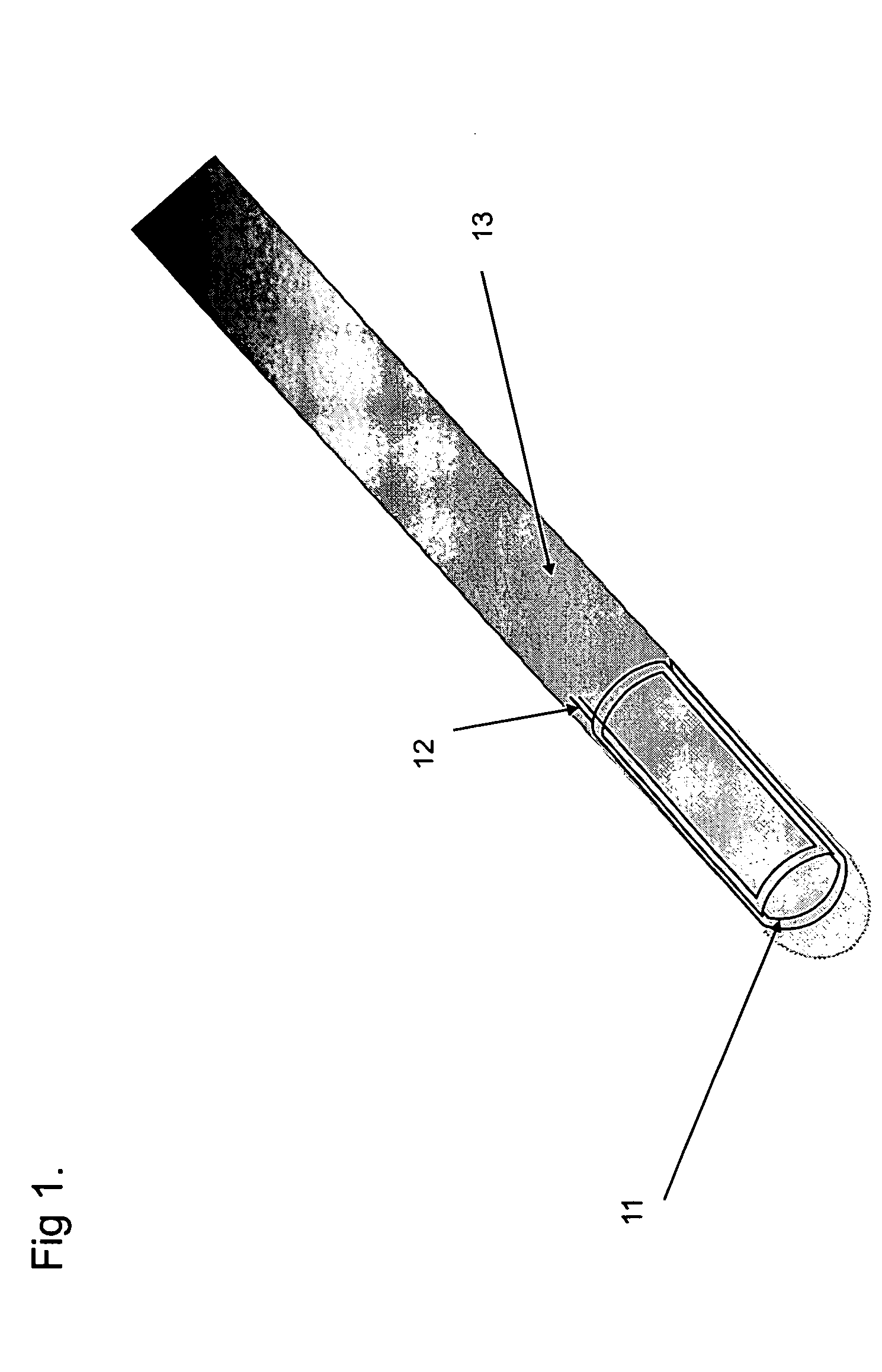
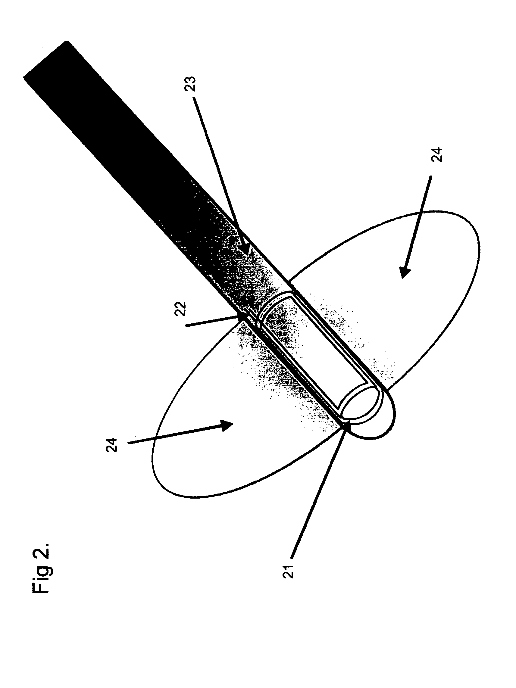
[0040] A technology is practiced including a medical device having a longitudinal axis, the device having at least one RF microcoil system in the device, at least one surface coil system around the regions (e.g., patient, organ, member, etc.) to be imaged, and a parallel imaging method support system (e.g., sufficiently advanced MR system as is known in the art for use in parallel imaging,, software presently available with commercial imaging systems for use in parallel imaging methods, and a viewing system (e.g., CRTY, plasma screen, hard copy media, LED display, etc.). The position of the at least one RF microcoil system and the at least one surface coil system with respect to the longitudinal axis and the patient define regions where different field volumes are provided by the at least one RF microcoil system and the at least one surface coil system. The medical device may have the surface microcoil system connected with at least one preamplifier in communication with a signal re...


PUM


Abstract

A method, system and apparatus provides a magnetic resonance (MR) responsive field of view within a volume of a patient. At least two radiofrequency (RF) surface coils are provided that at least in part MR responsively cover the volume, at least one MR responsive microcoil is provided within the volume, and MR responsive fields are simultaneously generated from the at least two RF surface coils and the at least one microcoil. The data of the RF responsive fields are then integrated with parallel imaging methods.

Description

BACKGROUND OF THE INVENTION [0001] 1. Field of the Invention [0002] The present invention relates to the field of medical devices, medical devices that are used within organisms, medical devices that are used with or incidental to Magnetic Resonance Imaging, or medical devices that are used with medical treatments after, during or preceding Magnetic Resonance Imaging of the region within which the treatment is planned. [0003] 2. Background of the Art [0004] Medical procedures may now be performed on areas of the patient which are relatively small. Procedures may be performed on small clusters of cells, within veins and arteries, and in remote sections of the body with minimally invasive techniques, such as without surgical opening of the body. As these procedures, such as balloon angioplasty, microsurgery, electrotherapy, and drug delivery are performed within the patient with minimally invasive techniques without major surgical opening of the patient, techniques have had to be deve...

Claims


Application Information

Patent Timeline
Patent Type & Authority Applications(United States)
IPC IPC(8): A61B5/05
CPCA61B5/055G01R33/287G01R33/34084G01R33/3415G01R33/5611
Inventor KUCHARCZYK, JOHNBRONSKILL, MICHAEL JOHN
Owner NEXGEN
Why Eureka
Try Eureka