Fluorescent probe and application thereof

A fluorescent probe, alkyl technology, applied in the field of analytical chemistry, can solve problems such as lack of biological samples, and achieve the effects of less damage to biological samples, good membrane permeability, and low cytotoxicity

Active Publication Date: 2018-11-13
INST OF CHEM CHINESE ACAD OF SCI
View PDF3 Cites 11 Cited by
  • Summary
  • Abstract
  • Description
  • Claims
  • Application Information

AI Technical Summary

Problems solved by technology

There are several fluorescent probes for labeling lysosomes on the market, such as LysoTracker Red, LysoTracker Green, etc., but there is still a lack of commercialized probes for labeling autolysosomes

Method used


Image

  • Fluorescent probe and application thereof
  • Fluorescent probe and application thereof
  • Fluorescent probe and application thereof

Examples


Embodiment 1

[0067] The fluorescent probe (1) and the LysoTracker Red probe of the embodiment of the present invention were used to perform fluorescence imaging on the cells, as follows:

[0068]

[0069] (1) Dissolve fluorescent probe (1) and LysoTracker Red probe with a small amount of DMSO;

[0070] (2) Add the two fluorescent probe solutions in step (1) to the culture medium respectively, and configure them as a culture solution containing a fluorescent dye 1 at a concentration of 10 μM and a culture solution of LysoTracker Red at a concentration of 1.0 μM;

[0071] (3) Use a pipette gun to pipette 1mL of the culture solution prepared in step (2), add it to the culture dish with HEK293 cells, and put it in 37°C, 5% CO 2 Cultivate in the incubator for 30min;

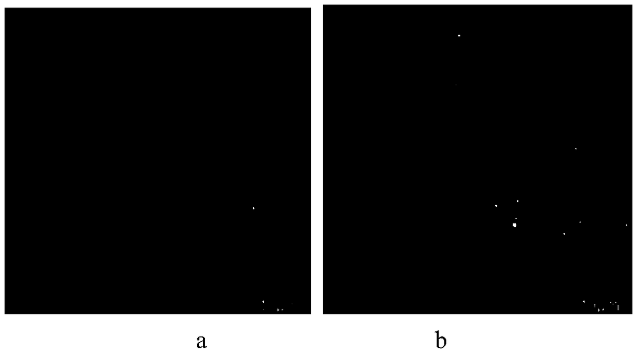
[0072] (4) The cultured cells were washed three times with PBS, and then 1 mL of blank mixed medium was added for fluorescent confocal imaging. The results were as follows: figure 1 As shown, wherein, a is a schematic diagram...

Embodiment 2

[0074] Fluorescence imaging of the process of cell starvation-induced lysosomal autophagy is monitored by using the fluorescent probe (2) of the embodiment of the present invention, specifically as follows:

[0075]

[0076] (1) Dissolve the fluorescent probe (2) with a small amount of ethanol;

[0077] (2) Add the fluorescent probe solution in step (1) to the culture medium containing 0, 5% and 10% embryonic bovine serum, and configure it as a culture solution containing fluorescent dye 2 at a concentration of 4 μM;

[0078] (3) Use a pipette gun to pipette 1 mL of the culture solution prepared in step (2) respectively, and add them to the culture dishes of human lung cancer cell A549 cultured with 0, 5% and 10% embryonic bovine serum medium, and place It is placed in 37 ℃, 5% CO 2 Cultivate in the incubator for 1h;

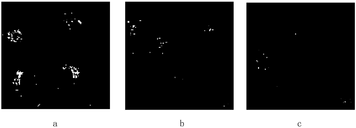
[0079] (4) The cultured cells were washed three times with PBS, and then 1 mL of blank mixed medium was added for fluorescent confocal imaging. The results...

Embodiment 3

[0081] The fluorescent probe (3) of the embodiment of the present invention is used to monitor the fluorescence imaging of the process of cell starvation-induced lysosomal autophagy, specifically as follows:

[0082]

[0083] (1) dissolve the fluorescent probe (3) with a small amount of methanol;

[0084] (2) adding the fluorescent probe solution in step (1) to a blank culture medium not containing embryonic bovine serum, and respectively configuring blank culture solutions containing fluorescent dye 3 at a concentration of 5 μM;

[0085] (3) Use a pipette gun to pipette 1 mL of the blank culture solution prepared in step (2) respectively, and add them to the culture dishes of cervical cancer HeLa cells that have been starved for 0, 0.5 and 1 hour respectively, and put them into At 37°C, 5% CO 2 Cultivate in the incubator for 20min;

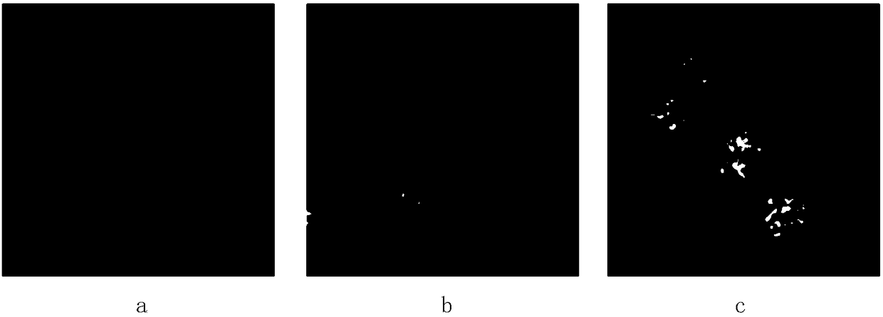
[0086] (4) The cultured cells were washed three times with PBS, and then 1 mL of blank mixed medium was added for fluorescent confocal imag...


PUM

PropertyMeasurementUnit
diameteraaaaaaaaaa

Abstract

The invention discloses a fluorescent probe and application thereof. The fluorescent probe is provided with a structure shown as a formula I or a stereoisomer of the fluorescent probe, wherein R1 is hydrogen, alkyls of C1 to C6, phenyl or alkyl-substituted phenyl; R2 to R9 are independent hydrogen atoms, alkyls of C1 to C6, halogenated alkyls of C1 to C6 and alkoxys or hydrogen atoms of C1 to C6;R10 and R11 are independent sulfo or sulfo-substituted alkyls of C1 to C6; X1 and X2 are independent carbon, oxygen, sulfur, selenium or tellurium. The fluorescent probe has the specific advantages ofgood biocompatibility, low cytotoxicity, small damage to a biological sample and good photobleaching resistance; long-time effective observation of a cell sample can be realized without influence from a pH value of a cell.

Description

technical field [0001] The present invention relates to the field of analytical chemistry, in particular to fluorescent probes and applications thereof, more specifically to fluorescent probes, the use of fluorescent probes in fluorescent imaging of intracellular autophagy-lysosomes and the A Method for Fluorescent Imaging of Enzymosomes. Background technique [0002] Lysosome is a very important organelle with a diameter of about 200-500nm, which exists in almost all eukaryotic cells. There are many enzymes and proteins in lysosomes, including acid hydrolase, trypsin and cathepsin, which are digestive organs in cells, which can decompose various proteins and other macromolecules, and have the function of dissolving or digesting. Autophagy lysosomes, also known as cytolysosomes, are formed by the fusion of primary lysosomes and vesicles containing endogenous substances from autophagy, or lysosomes engulfing the cytoplasm. Acts as a "scavenger" for the normal pathway of att...

Claims


Application Information

Patent Timeline
Patent Type & Authority Applications(China)
IPC IPC(8): C07D277/64C07D417/06C07D421/06C09K11/06G01N21/64
CPCC07D277/64C07D417/06C07D421/06C09K11/06C09K2211/1037C09K2211/104G01N21/6486
Inventor 孙红霞陈宏博唐亚林
Owner INST OF CHEM CHINESE ACAD OF SCI
Why Eureka
Try Eureka