Biocompatible tissue sealant for treatment of osteochondral and bone defects using an acellular matrix implant

a biocompatible tissue and bone technology, applied in the field of ace, can solve the problems of debilitating disability, affecting mobility, damage to the articular cartilage of active individuals and older generation adults,

Inactive Publication Date: 2006-04-20
KUSANAGI AKIHIKO +2
View PDF24 Cites 14 Cited by
  • Summary
  • Abstract
  • Description
  • Claims
  • Application Information

AI Technical Summary

Benefits of technology

[0027] Still another aspect of the current invention is a bone inducing composition or a carrier comprising said composition containing bone inducing agents such as a demineralized bone powder, calcium phosphate, hydroxyapatite, organoapatite, titanium oxide, poly-L-lactic or polyglycolic acid or a copolymer thereof or a bone morphogenic protein used in a method where the deposition of said composition into the bone lesion initiates migration of osteoblast and achieves natural healing of the underlying bone.
[0030] Still another aspect of the current invention is a bone inducing composition or a carrier comprising said composition containing bone inducing agents such as a demineralized bone powder, calcium phosphate, hydroxyapatite, organoapatite, titanium oxide, poly-L-lactic or polyglycolic acid or a copolymer thereof or a bone morphogenic protein alone, in combination, or incorporated into a carrier, such as a matrix, hydrogel, sponge, honeycomb, scaffold or a polymer of an aromatic organic acid, used in a method where the deposition of said composition into the bone lesion initiates migration of osteoblast and achieves natural healing of the underlying bone.
[0032] Yet another aspect of the current invention is a method for treatment of injured, damaged, diseased or aged articular cartilage using an acellular matrix implant implanted into a joint cartilage lesion in situ, said method further comprising a formation of a new superficial cartilage layer overgrowing and sealing the lesion in the joint articular cartilage by applying a top sealant over the lesion and further applying a bottom sealant over the bottom of the lesion, said bottom sealant providing protection of the lesion against a cell and blood debris migration.
[0037] b) preparation of a cartilage lesion for implantation of said implant, including a step of depositing a bottom sealant at the bottom of the cartilage lesion for sealing of said lesion and protecting the implant from migration of blood-borne agents;
[0045] Still another aspect of the current invention is an acellular matrix implant comprising a thermo-reversible gelation hydrogel (TRGH) deposited into a lesion cavity formed above the bottom sealant layer, or into the cavity between the top and bottom sealant, said TRGH deposited into said cavity either incorporated into a collagenous sponge or scaffold or as a sol at temperatures between about 5 to about 30° C., wherein within said cavity and at the body temperature said TRGH converts from the fluidic sol into a solid gel and in this form, its presence provides a structural support for migration of chondrocytes from a surrounding native cartilage and formation of extracellular matrix, wherein said TRGH is biodegradable, will disintegrate with time and be metabolically removed from the lesion and replaced with a hyaline cartilage.
[0053] Still yet another aspect of the current invention is a bone inducing composition or a carrier comprising said composition comprising bone inducing agents for treatment of osteochondral defects further in combination with an acellular matrix implant comprising a thermo-reversible gelation hydrogel (TRGH) each deposited separately into a bone or cartilage lesion, wherein said composition provides a means for rebuilding the bone and migration of osteoblast into the bone lesion and wherein said implant provides a structural support for migration of chondrocytes from a surrounding native cartilage and formation of extracellular matrix.

Problems solved by technology

Damage to the articular cartilage which occurs in active individuals and older generation adults as a result of either acute or repetitive traumatic injury or aging is quite common.
Such damaged cartilage leads to pain, affects mobility and results in debilitating disability.
For example, severe and chronic forms of knee joint cartilage damage can lead to greater deterioration of the joint cartilage and may eventually lead to a total knee joint replacement.
Osteochondral diseases or injuries, which are a combination lesions of bone and cartilage, present yet another challenge for a treatment of which need is not being met by the currently available procedures and methods.

Method used


Image

  • Biocompatible tissue sealant for treatment of osteochondral and bone defects using an acellular matrix implant
  • Biocompatible tissue sealant for treatment of osteochondral and bone defects using an acellular matrix implant
  • Biocompatible tissue sealant for treatment of osteochondral and bone defects using an acellular matrix implant

Examples


example 1

Preparation of Acellular Collagenous Implants

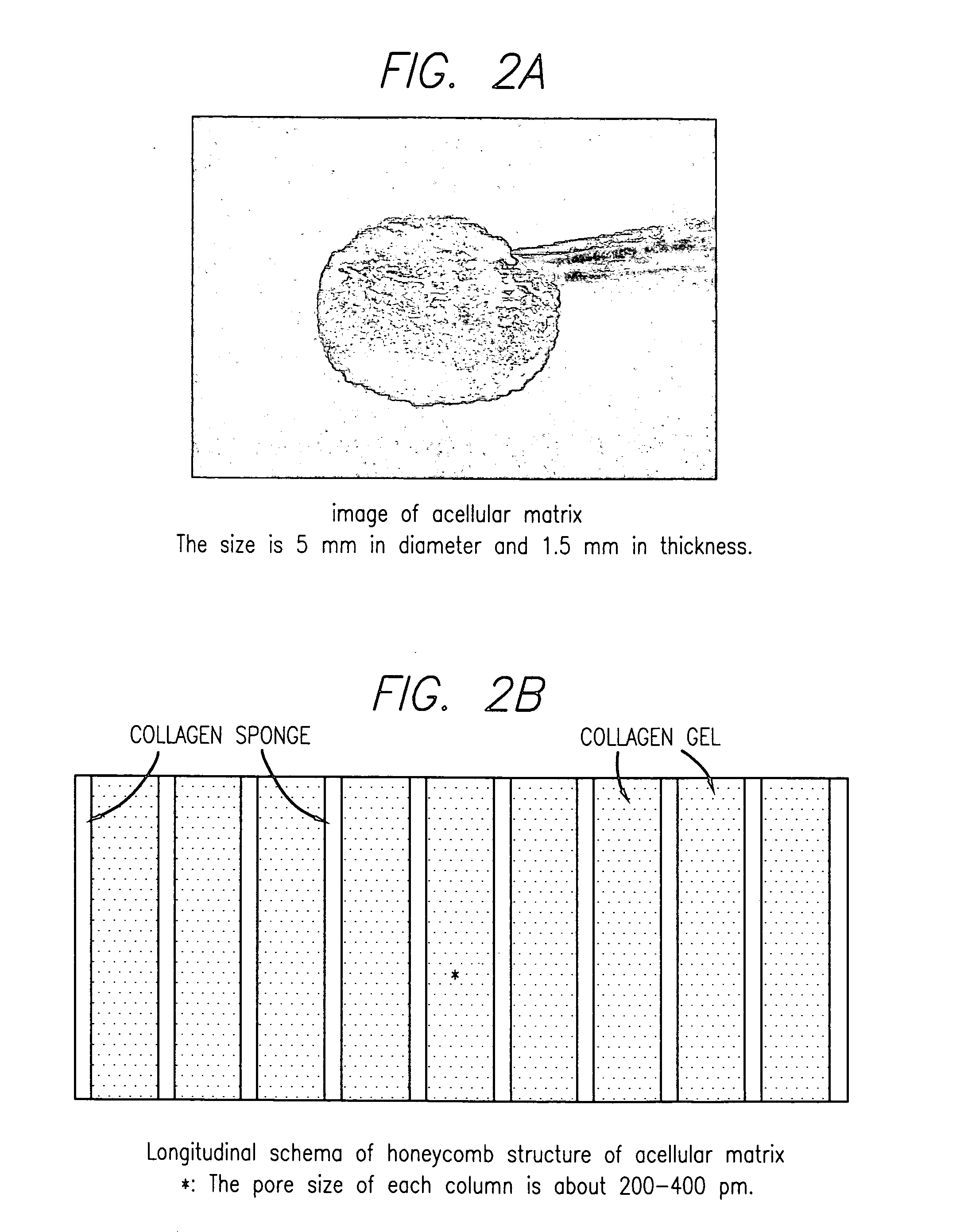
[0383] This example illustrates preparation of the acellular matrix implant.

[0384] 300 grams of a 1% aqueous atelocollagen solution (VITROGEN®), maintained at pH 3.0, is poured into a 10×20 cm tray. This tray is then placed in a 5 liter container. A 50 ml open container containing 30 ml of a 3% aqueous ammonia solution is then placed next to the tray, in the 5 liter chamber, containing 300 grams of said 1% aqueous solution of atelocollagen. The 5 liter container containing the open trays of atelocollagen and ammonia is then sealed and left to stand at room temperature for 12 hours. During this period the ammonia gas, released from the open container of aqueous ammonia and confined within the sealed 5 liter container, is reacted with the aqueous atelocollagen resulting in gelling said aqueous solution of atelocollagen.

[0385] The collagenous gel is then washed with water overnight and, subsequently, freeze-dried to yield a sponge like ma...

example 2

Biochemical and Histological Assays

[0391] This example describes assays used for biochemical and histological studies.

[0392] For biochemical (DMB) assay, the implant taken from the animal after certain time following the implantation, transferred to microcentrifuge tubes and digested in 300 μl of papain (125 μg / ml in 0.1 M sodium phosphate, 5 mM disodium EDTA, and 5 mM L-cysteine-HCl) for 18 hours at 60° C. S-GAG production in the implant is measured using a modified dimethylene blue (DMB) microassay with shark chondroitin sulfate as a control according to Connective Tissue Research, 9: 247-248 (1982).

[0393] DNA content is determined by Hoechst 33258 dye method according to Anal. Biochem., 174:168-176 (1988).

[0394] For histological assay, the remaining implants from each group were fixed in 4% paraformaldehyde. The implants were processed and embedded in paraffin. 10 μm sections were cut on a microtome and stained with Safranin-O (Saf O).

[0395] For immunohistochemistry, the sam...

example 3

Evaluation of Integration of Acellular Matrix Implant in a Swine Model

[0396] This example describe the procedure and results of study performed for evaluation of integration of porcine in a swine model.

[0397] An open arthrotomy of the right knee joint was performed on all animals, and a biopsy of the cartilage was obtained.

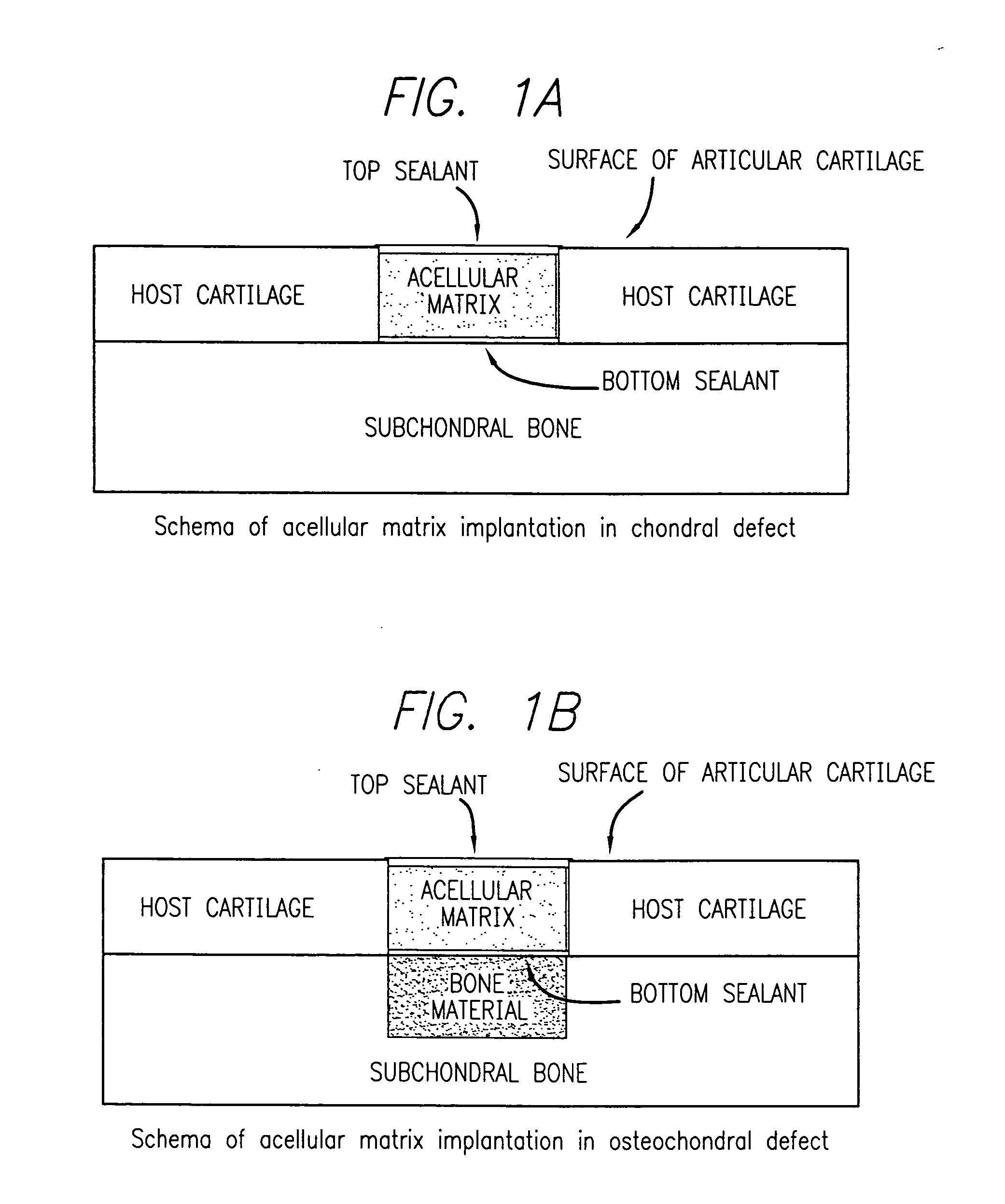
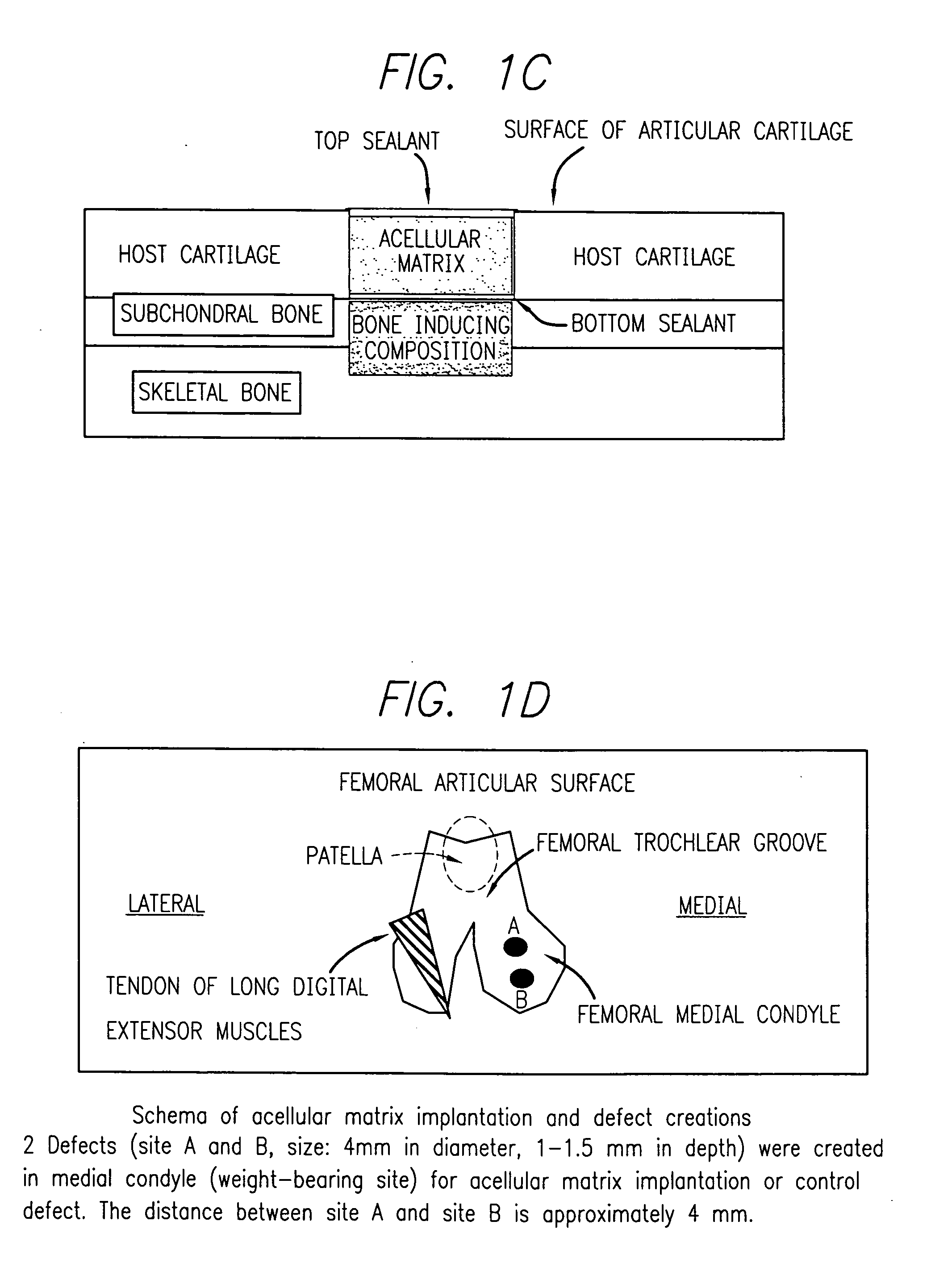
[0398] A defect was created in the medial femoral condyle of the pig's right knee. This defect (control) was not implanted with an acellular matrix implant but was left intact. Following surgery, the joint was immobilized with an external fixation implant for a period of about two weeks. Two weeks after the arthrotomy on the right knee was performed, an open arthrotomy was performed on the left knee and defects were created in this medial femoral condyle. The acellular matrix implant was implanted within the defect (s) in this knee which was similarly immobilized. The operated sites were subsequently viewed via arthroscopy two weeks after implantation or defect...


PUM

PropertyMeasurementUnit
bond strengthaaaaaaaaaa
tensile strengthaaaaaaaaaa
peel strengthsaaaaaaaaaa

Abstract

An acellular matrix implant for treatment of defects and injuries of articular cartilage, bone or osteochondral bone and a method for treatment of injured, damaged, diseased or aged articular cartilage or bone, using the acellular matrix implant implanted into a joint cartilage lesion in situ and a bone-inducing composition implanted into an osteochondral or bone defect. A method for repair and restoration of the injured, damaged, diseased or aged cartilage or bone into its full functionality by implanting the acellular matrix implant between two layers of biologically acceptable sealants and / or the bone-inducing composition into the osteochondral bone or skeletal bone defect. A method for fabrication of the acellular matrix implant of the invention. A method for preparation of bone-inducing composition.

Description

[0001] This application is based on and claims priority of the Provisional Application Ser. No. 60 / 496,971, filed Aug. 20, 2003, which is incorporated herein by reference.BACKGROUND OF THE INVENTION Field of Invention [0002] The current invention concerns acellular matrix implants and compositions for treatment of articular cartilage, bone or osteochondral defects and injuries and a method for treatment of such osteochondral defects and / or injured, damaged, diseased or aged articular cartilage or bone using an acellular matrix implant implanted into a joint cartilage lesion and / or into the osteochondral defect in situ wherein the osteochondral or bone defect is further implanted with a bone inducing composition or a carrier comprising said composition. The acellular matrix implant of the invention comprises a two or three dimensional biodegradable scaffold structure implanted into the joint cartilage lesion typically below or over one, two or several layers, or between two layers of...

Claims


Application Information

Patent Timeline
Patent Type & Authority Applications(United States)
IPC IPC(8): A61K38/43A61F13/00A61F2/00A61F2/30A61L27/24A61L27/34A61L27/36A61L27/52A61L27/58
CPCA61F2/30756A61F2002/30535A61F2250/0058A61L27/24A61L27/34A61L27/3633A61L27/3654A61L27/52A61L27/58A61L2430/06A61L27/3608
Inventor KUSANAGI, AKIHIKOTARRANT, LAURENCE J. B.SCHMIDT, MARY BETH
Owner KUSANAGI AKIHIKO
Why Eureka
Try Eureka