Tissue-adhesive formulations

a technology of tissue adhesive and formulation, applied in the field of tissue adhesive formulation, can solve the problems of limited life of formulation, poor initial adhesion, and difficult application to the desired area of tissue, and achieve the effects of reducing or eliminating cracking and crumbling of tissue-reactive materials, good initial adhesion, and maintaining pliability and physical properties of suppor

Inactive Publication Date: 2006-05-18
TISSUEMED LTD
View PDF99 Cites 75 Cited by
  • Summary
  • Abstract
  • Description
  • Claims
  • Application Information

AI Technical Summary

Benefits of technology

[0008] The formulation according to the invention is advantageous primarily in that it can be easily applied to a tissue surface using a simple applicator or delivery device. As it is applied in solid form, the particulate formulation adheres to the tissue surface and does not spread unduly. The formulation exhibits good initial adhesion to the tissue surface, this being believed to be due to van der Waals forces and / or hydrogen bonding between the formulation and the tissue surface. On contact with the tissue surface the formulation becomes hydrated, thereby causing reaction between the tissue-reactive functional groups and the underlying tissue surface. Such reactions between the tissue-reactive functional groups and the underlying tissue results in high adhesion between the formulation and the tissue surface, and hence between tissues hat are joined using the adhesive formulation. Reaction may also take place between the tissue-reactive functional groups and the other components of the formulation to form a strong, flexible and tissue-adherent gel. This formulation thus absorbs physiological fluids (as a consequence of application onto exuding tissue surfaces), and any additional solutions used to hydrate the formulation following application (such fluids can be commonly used solutions used in surgical irrigation), becoming gelatinous and adherent to the tissue surfaces, and thereby providing an adhesive sealant, haemostatic and pneumostatic function.
[0009] In addition, because the formulation is made up in solid form that is, until hydrated by contact with the tissue surface (and subsequent hydration), essentially inactive, the formulation is not prone to premature reaction and as a result its shelf-life may be considerable, eg more than six months when stored at room temperature and stored appropriately. This further enables the formulation to be packaged in relatively large quantities that can be dispensed and used over a considerable time period, without the risk of substantial wastage.
[0012] The sheet according to the invention is advantageous primarily in that it bonds effectively to tissue, enabling it to be used in a variety of medical applications. The invention enables coating of the tissue-reactive materials onto (and into) a three-dimensional structural support, whilst maintaining the pliability and physical properties of the support. Furthermore, the adhesive performance of the tissue-reactive materials is not compromised when delivered to the target tissue in this form. Where, as in preferred embodiments, the support is perforated, the perforations provide a means of anchoring the tissue-reactive materials in the support. This reduces or eliminates cracking and crumbling of the tissue-reactive material as it is applied to the support (core), which would result in sub-optimal coverage of the target tissue, and thereby compromise the adhesive / sealant effects of the sheet.
[0013] The sheet exhibits good initial adhesion to the tissue to which it is applied (and may thus be described as “self-adhesive”), and furthermore remains well-adhered to the tissue over a longer timescale. Without wishing to be bound by any theory, it is believed that the initial adhesion of the sheet to the tissue is attributable to electronic bonding of the sheet to the tissue, and this is supplemented or replaced by chemical bonding between the tissue-reactive functional groups of the formulation and the tissue, in particular between amine and / or thiol groups on the tissue surface and the tissue-reactive groups of the sheet. Where the structural inner core of the device is perforated, and is coated on both sides with the tissue-adhesive formulation, the perforations facilitate hydration and cross-linking of the formulation on both sides of the core such that the core becomes enclosed within a three-dimensional matrix of cross-linked material.
[0014] The use of the sheet reduces or eliminates the need for additional means of mechanical attachment to the tissue (eg sutures or staples), or the need to provide external energy in the form of heat or light to bring about adherence of the sheet to the underlying tissue. Another advantage of the sheet according to the invention is that it is applied to the tissue as a preformed article, rather than being prepared by mixing of materials immediately prior to use.

Problems solved by technology

If the formulation is of low viscosity, it may spread from the area of application and hence be difficult to apply precisely to the desired area of tissue.
If the formulation is more viscous, on the other hand, it may be difficult to dispense.
In either case, the formulation, being prepared in hydrated form, may have a limited lifetime and may be subject to premature curing.
Also, the preparation of formulations immediately prior to use by mixing of ingredients is obviously laborious and time-consuming.
In addition to these drawbacks, the degree of adhesion between tissue surfaces that is provided by such formulations may be less than would be desired.
A disadvantage of products proposed hitherto, however, is that the degree of adhesion to the underlying tissue, particularly in the longer term, may be inadequate.
In addition, the flexibility of the product may be insufficient for it to conform readily to the surface to which it is applied, which may also have an adverse effect on its adhesion.
As a result of the inadequate adhesion of these products, it may be necessary to provide further reinforcement, eg through mechanical attachment using sutures, staples or the like.
Clearly, such approaches introduce further drawbacks.
In many instances, the use of such fastenings is either not wholly effective (eg on the lung) or undesirable, as their introduction gives rise to further areas of tissue weakness.
Such energy sources may be expensive and difficult to operate, particularly in the confines of an operating theatre or similar environment.
Also, the use of external energy for attachment can be both time-consuming and (in some cases) requires significant careful judgement on the part of the surgeon, to evaluate when sufficient energy has been delivered to effect attachment without damaging the underlying tissue.

Method used


Image

  • Tissue-adhesive formulations
  • Tissue-adhesive formulations
  • Tissue-adhesive formulations

Examples


example 1

Synthesis of NHS-activated PVP-co-PAA

(a) Polymerisation of Acrylic Acid and N-vinyl-2-pyrrolidone

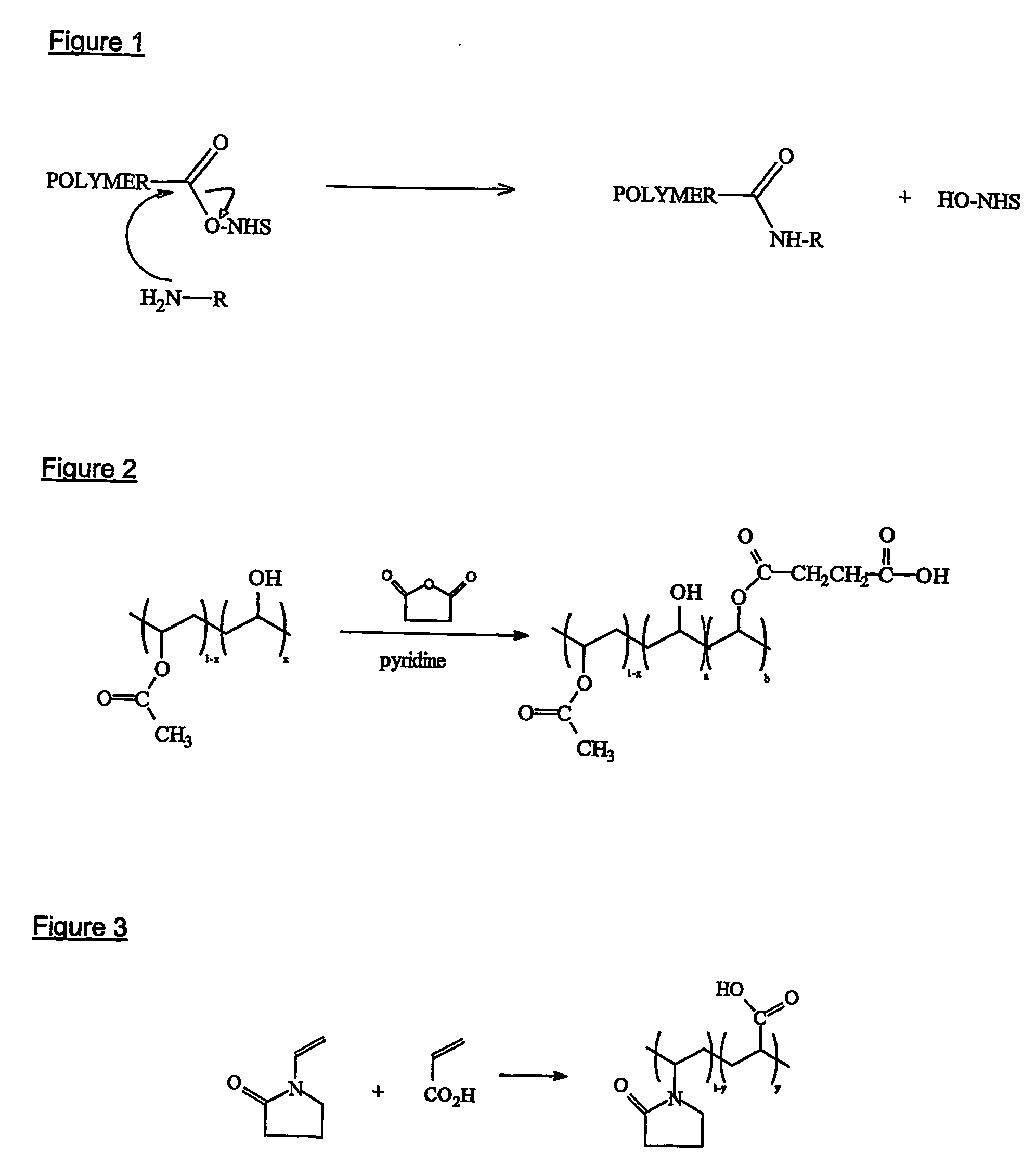
[0151] The polymer is formed via the polymerisation of monomers such as N-vinyl-2-pyrrolidone and acrylic acid, as shown in FIG. 3.

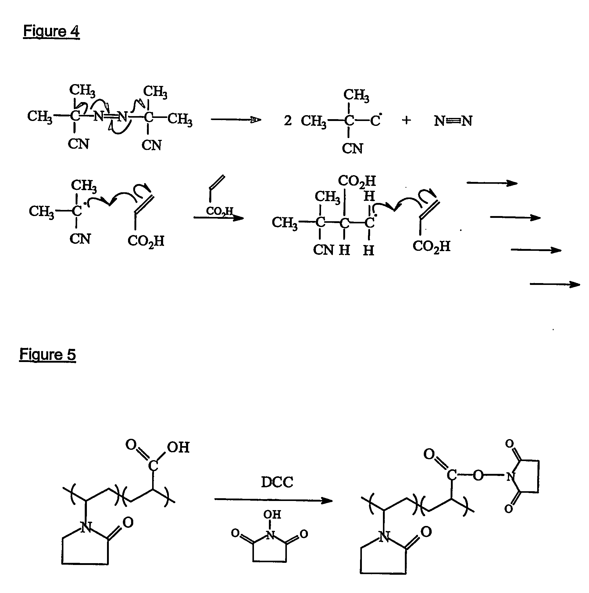
[0152] A number of methods may be used to initiate the polymerisation, such as free radical, ionic (cationic or anionic), thermal, UV, redox etc. Free radical polymerisation is the preferred polymerisation method and 2-2′-azo-bis-isobutyrynitrile (AIBN) is the preferred initiator. The AIBN decomposes into two radicals which can then attack the carbon-carbon double bond in the vinylic monomer (acrylic acid) as shown in FIG. 4.

[0153] This will continue until termination of chain growth, via combination, disproportionation etc.

[0154] The reaction solvent may be N,N′-dimethylformamide, toluene, or any other suitable solvent with a boiling point greater than 100° C. Toluene is the currently preferred solvent.

[0155] A typical polymerisation method is as foll...

example 2

Alternative Synthesis of NHS-Activated PVP-co-PAA

(a) Polymerisation

[0162] 400ml of dried toluene is heated to 80±2° C. in a round bottomed flask using an oil bath or isomantle. Oxygen is removed from the solvent by bubbling oxygen-free nitrogen through the toluene for at least 30 minutes. 0.1 g (0.006 moles) of azo-iso-butyronitrile (AIBN) dissolved in 2 ml of toluene is added to the reaction flask using a syringe, immediately followed by 45.02 g (0.406 moles) of 1-vinyl-2-pyrrolidone and 7.02 g (0.092 moles) of acrylic acid. The reaction is left under nitrogen at 80±2° C. for 17 hours; the polymer is insoluble in toluene and forms a white precipitate as the reaction proceeds. After 17 hours, a further 0.1 g (0.006 moles) of AIBN is added and the reaction is kept at 80±2° C. for one further hour to polymerise any remaining monomer. The polymer is isolated by pouring into 2000 ml of rapidly stirred 1:1 hexane:diethyl ether and subsequent filtration using a 10-16 μm filter. The pol...

example 3

Blending of NHS-Activated PVP-co-PAA with Freeze-Dried Albumin

[0165] a) Powders of NHS-activated PVP80-co-PAA20 copolymers (ie copolymers consisting of 80 mol % vinyl pyrrolidone-derived units and 20 mol % acrylic acid-derived units) have been blended (in ratios of 1:1, 2:1 and 4:1) with freeze-dried porcine albumin (Sigma Aldrich; previously buffered to pH 10.5).

b) Powders of NHS-activated PVP70-co-PAA30 copolymers (70 mol % vinyl pyrrolidone: 30mol % acrylic acid) have been blended (1:1) with freeze-dried human albumin (Baxter human albumin solution (20%) previously buffered to pH 10.5).

c) Powders of NHS-activated PVP70co-PAA30 copolymers have been blended (2:1) with freeze-dried porcine albumin (previously buffered to pH 10.5).


PUM

PropertyMeasurementUnit
molar ratioaaaaaaaaaa

Abstract

A tissue-adhesive formulation consists of a naturally occurring or synthetic polymerisable and / or cross-linkable material in particulate form, the polymerisable and / or cross-linkable material being in admixture with particulate material comprising tissue-reactive functional groups. The formulation may be used in the preparation of a tissue-adhesive sheet, by applying the formulation to at least one side of a core of a naturally occurring or synthetic polymeric material.

Description

FIELD OF THE INVENTION [0001] This invention relates to materials suitable for use as tissue adhesives and sealants, and to a flexible multilamellar sheet, patch or film comprising such materials for topical application to internal and external surfaces of the body, for therapeutic purposes. The invention also relates to a process for the preparation of such products, and to methods of using such products. In particular the invention relates to materials that are formulated as loose or compacted powders and to a self-adhesive, biocompatible and hydratable polymeric sheet with such materials applied to a suitable support, which may be used for therapeutic purposes such as wound healing, joining, sealing and reinforcing weakened tissue, and for drug delivery, and to a process for preparing, and methods of using, such a sheet. BACKGROUND OF THE INVENTION [0002] There is considerable interest in the use, for a number of surgical or other therapeutic applications, of materials that adher...

Claims


Application Information

Patent Timeline
Patent Type & Authority Applications(United States)
IPC IPC(8): A61K9/70A61L15/00A61L15/22A61L15/32A61L24/04A61L24/10A61L31/04
CPCA61L15/225A61L15/32A61L24/043A61L24/108A61L31/041A61L31/047A61L24/04A61L24/10
Inventor FORTUNE, DAVID HARRYKETTLEWELL, GRAEMEMANDLEY, DAVID JOHNTHOMPSON, IANCOOK, DIANE
Owner TISSUEMED LTD
Why Eureka
Try Eureka