
Convective flow tissue assembly

a tissue and convective flow technology, applied in the field of systems and methods for producing tissue constructs in vitro, can solve the problems of increasing the burst strength of grafts, invasive graft harvesting, and patients often lack adequate autogenous vessels to serve as bypass conduits, etc., to achieve accurate tissue morphology, avoid delamination and uniformity issues, and maintain differentiation phenotyp

Inactive Publication Date: 2006-05-18
LA JOLLA BIOENG INST
View PDF10 Cites 4 Cited by
  • Summary
  • Abstract
  • Description
  • Claims
  • Application Information

AI Technical Summary

Benefits of technology

"The present invention provides a new way to create tissues in the laboratory using a convective flow of culture medium. This method involves assembling cells on a porous mandrel using drag forces, which is then removed to form the final tissue construct. This approach results in tissues that are completely biological, have accurate tissue morphology, and maintain their differentiated phenotype and mechanical properties. The method also ensures proper nutrient and oxygen supply to all parts of the tissue. The use of adult, autologous cells and a convective flow in a bioreactor makes the method suitable for both FDA approval and commercialization. The invention is also automated and scalable, making it suitable for large-scale production. Overall, the invention provides an optimal and effective way to produce tissues in the laboratory."

Problems solved by technology

Unfortunately, not only is graft harvesting invasive but patients frequently lack adequate autogenous vessels to serve as bypass conduits, particularly in the case of patients who require repeated, multiple bypass procedures.
Unfortunately, their grafts displayed burst strengths of less than 10 mm Hg and even the use of DACRON sleeves did not increase the burst strength of the grafts beyond 350 mm Hg.
While these vessels acted as permeability barriers, the poor mechanical properties and the requirement of artificial materials limited this approach.
Unfortunately, the maximum demonstrated burst strength of these engineered tissues has been only 225 mm Hg, which is much lower than the 1700 mm Hg burst strength of human saphenous veins.
Thus, it does not seem likely that this approach will see successful clinical application.
Other approaches have also been developed, but these approaches also have various disadvantages and insufficiencies.
Unfortunately, the in vivo animal study showed mixed results.
Furthermore, problems with delamination of the layers and transport insufficiency have been observed.
Finally, this approach utilized neonatal cells and the labor-intensive methodology required to use neonatal cells is impractical for large scale production of TEVGs having consistent biological and mechanical properties.
While the initial results were very promising for tissues engineered with this technique, these TEVGs were not 100% biological and the degradation of the PGA mesh appeared to affect the SMCs, dedifferentiating them, perhaps due to the local hyperacid conditions.
Unfortunately, the mechanical properties of the vessels were not characterized.
Furthermore, the assembly methodology was invasive and relied on the silastic mandrel producing a strong foreign body inflammatory response.
As such, this method is not well suited for producing grafts for sick patients and is not likely to see approval by the US Food and Drug Administration for wide spread clinical use.
It is quite clear that despite their scientific value, all of the previous studies described above and known in the art have limitations regarding their ability to achieve clinical efficacy and practical application for disease management.

Method used


Image

  • Convective flow tissue assembly
  • Convective flow tissue assembly
  • Convective flow tissue assembly

Examples


Embodiment Construction

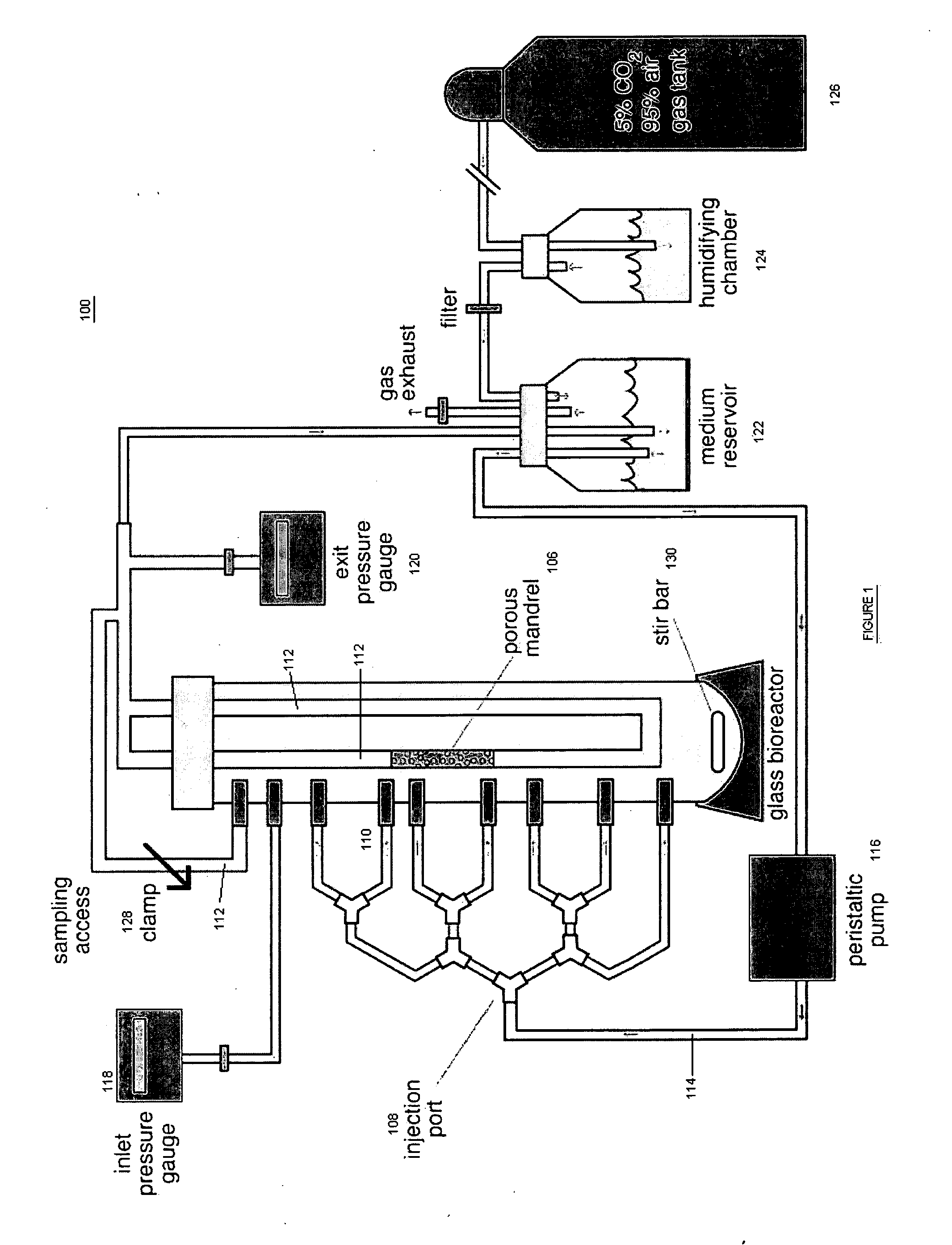
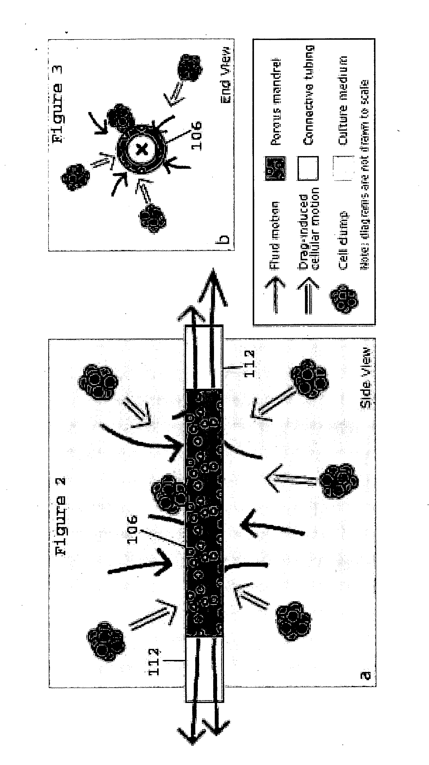
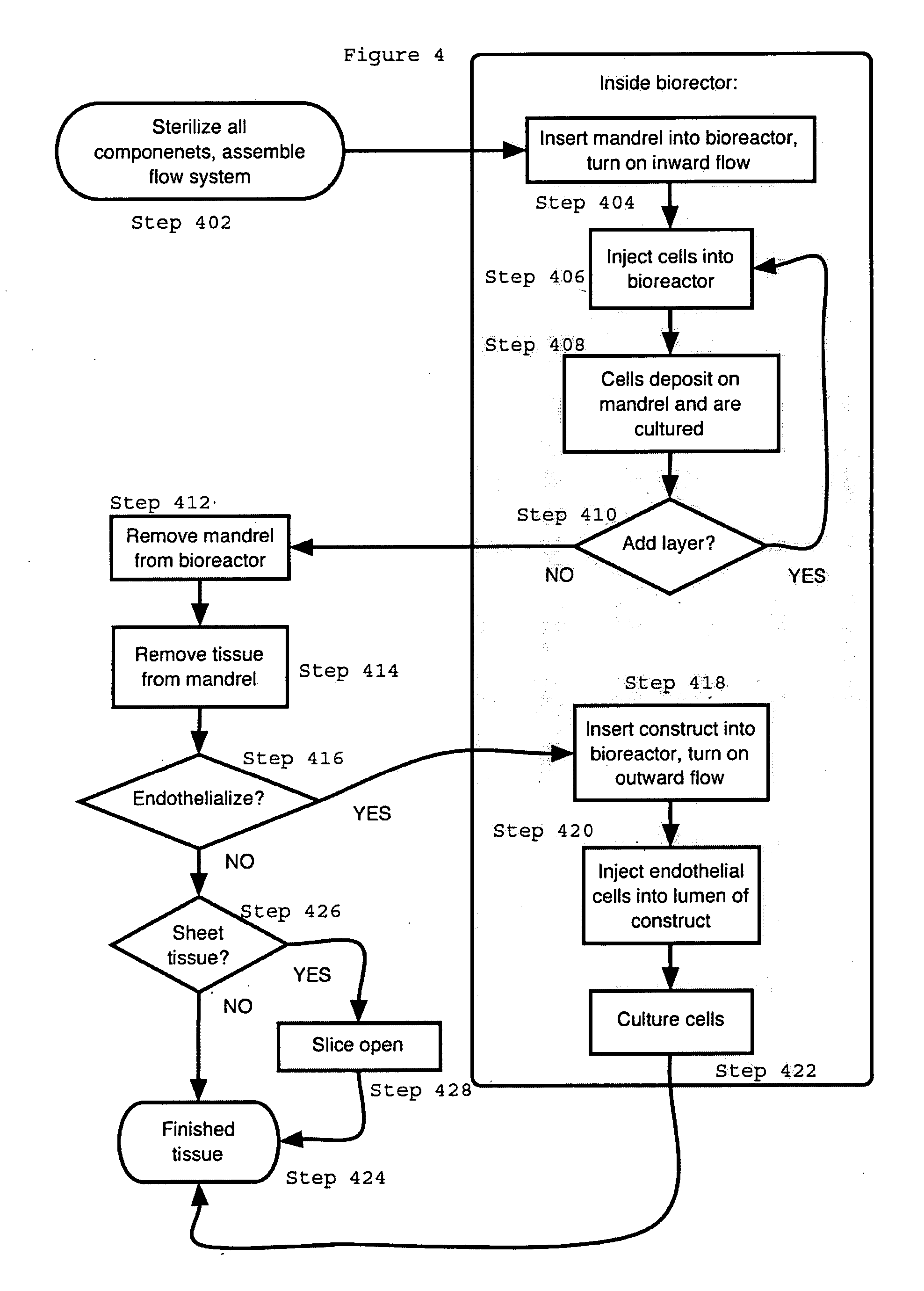
[0018] The present invention provides for novel systems and methods for performing convective flow tissue assembly and producing tissue constructs in vitro. The processes of the present invention generally involve the assembling of cells on an inert, porous mandrel, which only serves as a template and will ultimately be removed, via drag forces produced by a radial, convective flow of culture medium. The convective flow may be maintained by holding the luminal side of the mandrel at a lower pressure than that of the main bioreactor chamber, resulting in a transmural pressure gradient and radial flow within the bioreactor. As a result, cells may be actively deposited on the mandrel by fluid drag forces, thereby maximizing efficiency and ensuring a uniform distribution of cells on the mandrel. The pressure gradient will be radially symmetric and will result in fluid flow, which will also be symmetric in the radial direction.

[0019] The CFTA cell deposition system of the present invent...


PUM

PropertyMeasurementUnit
wall shear stressaaaaaaaaaa
pressureaaaaaaaaaa

Abstract

The present invention provides for an improved in vitro tissue assembly system and related methods that includes and uses a bioreactor, a porous mandrel disposed in the bioreactor, and components that provide for the circulation of culture media and cell suspensions within the bioreactor and through the porous mandrel. The circulation of the culture media and cell suspensions within the bioreactor produces a radial, convective flow and drag forces that result in the deposition of cells on the mandrel to form a tissue construct. Upon completion of the culture and tissue formation process, the tissue construct may be removed from the mandrel for subsequent in vivo use.

Description

FIELD OF THE INVENTION [0001] The present invention relates to systems and methods for producing tissue constructs in vitro. BACKGROUND [0002] Heart disease is the leading cause of death in the United States, affecting 12 million Americans and with an annual economic burden that exceeds 110 billion dollars. Coronary heart disease accounts for the largest portion of heart disease cases. Presently, coronary artery bypass graft (“CABG”) is an accepted approach both on an elective and emergency basis for restoring blood flow to areas of the heart affected by coronary artery stenosis. CABG is one of the most common medical procedures with more than 600,000 conducted annually. [0003] Clinically, in a CABG procedure the stenotic coronary artery is bypassed using a graft consisting of a saphenous vein or a mammary artery conduit. Unfortunately, not only is graft harvesting invasive but patients frequently lack adequate autogenous vessels to serve as bypass conduits, particularly in the case...

Claims


Application Information

Patent Timeline
Patent Type & Authority Applications(United States)
IPC IPC(8): C12N5/08
CPCC12M21/08C12M25/02
Inventor FRANGOS, JOHN A.SOBOLEWSKI, PETER
Owner LA JOLLA BIOENG INST
Learn More